
- 740 pages
- English
- ePUB (mobile friendly)
- Available on iOS & Android
eBook - ePub
About this book
The book has two intentions. First, it assembles the latest research in the field of medical imaging technology in one place. Detailed descriptions of current state-of-the-art medical imaging systems (comprised of x-ray CT, MRI, ultrasound, and nuclear medicine) and data processing techniques are discussed. Information is provided that will give interested engineers and scientists a solid foundation from which to build with additional resources. Secondly, it exposes the reader to myriad applications that medical imaging technology has enabled.
Tools to learn more effectively

Saving Books

Keyword Search

Annotating Text

Listen to it instead
Information
1
Future of Medical Imaging
Mark Nadeski and Gene Frantz
CONTENTS
1.1 Introduction
1.2 Where Are We Going?
1.2.1 EyeCam
1.3 Making Health Care More Personal
1.3.1 Advances in Digital and Medical Imaging
1.4 How Telecommunications Complements Medical Imaging
1.5 Automated Monitoring
1.6 Future of Technology
1.6.1 Remembering Our Focus
1.7 What We Can Expect from Technology
1.7.1 Development Cost
1.7.2 Performance
1.7.3 Multiprocessor Complexity
1.7.3.1 Multiprocessing Elements
1.7.4 Power Dissipation
1.7.4.1 Lower Power into the Future
1.7.4.2 Perpetual Devices
1.8 Integration through SoC and SiP
1.9 Defining the Future
References
1.1 Introduction
There are those who fear technology is nearly at the physical limitations of our understanding of nature, so where can we possibly go from here? But technology is not where our limits lie. Integrated circuits (ICs) have always exceeded our ability to fully utilize the capacity they make available to us, and the future will be no exception, for technology does not drive innovation. In truth, it is innovation and human imagination which drive technology.
1.2 Where Are We Going?
The broad field of medical imaging has seen some truly spectacular advances in the past half-century, many that most of us take for granted. Once a marvel only in the laboratory, advances such as real-time and Doppler ultra-sonography, functional nuclear medicine, computed tomography, magnetic resonance imaging, and interventional angiography have all become available in clinical settings.
It is easy to sit back in wonder at how far the field of medical imaging has come. However, in this chapter, we glimpse into the future. Some of this future is quickly taking shape today, though some of it will not arrive for years, if not decades.
Specifically, we look at how advances in medical imaging are based on existing technology; how these technologies will provide more capacity and capabilities than we can conceivably exploit; and finally lead in to the conclusion that the future of medicine is not limited by what we know, but rather by what we can imagine.
Let us begin by looking at the edge of what is real, that wonderful place where ideas are transformed into reality.
1.2.1 EyeCam
For centuries, humanity has dreamed of being able to make the blind see. And, for as long, restoring a person's eyesight has been considered a feat commonly categorized as “a miracle.”
About 10 years ago, Texas Instruments (TI) began collaborating with a medical team at Johns Hopkins, well known for its ability to make miracles happen. The team's goal was to develop a way to take the signal from a camera and turn it into electrical impulses that could then be used to excite the retina as shown in Figure 1.1. If successful, they could return some level of vision back to individuals who had lost their eyesight due to retinitis pig-matosa, a disease that affects more than 100,000 people in the United States alone.*
Now at the University of Southern California, this team continues to make significant progress. The project has evolved considerably over the years. Its initial conception consisted of mounting a camera on a pair of glasses that would require patients to rotate their heads in order to look around. Today, the team is working to actually implant a camera module within the eye since it is much more natural to let the eye do the moving to point the camera in the right direction. However, it is one thing to say, implanting a camera in a person's eye is more practical than mounting it on glasses and quite another to achieve it. A number of challenges come to mind:
* Source: http://www.wrongdiagnosis.com/r/retinitis_pigmentosa/stats-country.htm
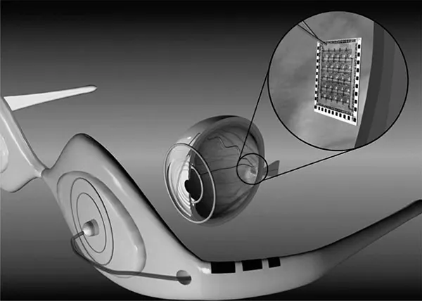
FIGURE 1.1
(See color insert.)
Example of the eyecam created and tested at the University of Southern California.
(See color insert.)
Example of the eyecam created and tested at the University of Southern California.
Size: The complete camera module has to be significantly smaller than an eyeball in order to fit.
Power: The camera must have exceptionally low power consumption. At the very least, the energy needs to be scavenged from body heat, the surrounding environment, or a yet-to-be-invented wireless power circuit.
Heat: Initial cameras may rely upon a connected power source. Even so, it is critical that the camera should not produce much heat. To be practical, the camera must be able to dissipate enough power so as not to heat the eye to the point of discomfort.
Durability: The camera must be packaged in such a way as to be protected from the fluids in the eye.
Currently working with Georgia Tech University and experts at TI, the team at USC is busy making all this happen. Is such an ambitious project even possible? Although success has yet to be seen, the team envisions a successful completion of the project. And they have good reason to be confident for they are only just pressing at the edges of possibility.
Much of what lies ahead of us in the world of medicine is the identification of technologies and devices from other parts of our world that we can apply to medical electronics. For example, Prof. Armand R. Tanguay, Jr., Principal Investigator on the “eyecam” project, acknowledges that they have many ideas about where else in the body they could implant a camera.*
Here a camera, there a camera,
In the eye a little camera.
Old Doc Donald had a patient.
E, I, E, I, O.
Certainly, there is more than one verse to this song. The question we might ask ourselves is what do we imagine we need next?
1.3 Making Health Care More Personal
A device that can help the blind to see is a life-changing application of medical technology. Not all medical devices will have such a dramatic effect on the way we live. Most of the changes in medical care will have a much lower profile, for they will be incorporated into our daily lives. However, while their application may be more subtle, the end result will certainly still be quite profound.
The future of medicine is based upon a firm foundation of existing technologies. What is new, in many cases, is not the technology itself, but rather how the technology is applied in new ways. Consider these key technologies:
- Digital imaging
- Telecommunications
- Automated monitoring
Each of these technologies is already firmly established in a number of disparate industries. Specifically applying them to medicine will still require creativity and hard work, but doing so will enable entirely new applications. Perhaps most importantly, for health care providers and their patients, the resulting advances will help shift health care into becoming a more routine part of daily life, creating a future where medical devices help us
- Manage our chronic conditions
- Predict our catastrophic diseases
- Enable us to live out our final months/years in the comfort of our homes
* Unfortunately, we cannot print any of these exciting ideas without a nondisclosure agreement in place.
1.3.1 Advances in Digital and Medical Imaging
Improving health care is the ultimate goal behind advances in medicine. As medical imaging advances, it will allow patients to have more personalized and targeted health care. Imaging, diagnosis, and treatment plans will continue to become more specialized and customized to a patient's particular needs and anatomy. We may even see therapies that are tailored to a person's specific genetics. Look at how far we have come already:
The migration to digital files: Photographic plates were once used to “catch” x-ray images. These plates gave way to film, which in turn are now giving way to digital radiography. Through the use of advanced digital signal processing, x-ray signals now can be converted to digital images at the point of acquisition while imposing no loss in image clarity. Digital files have a variety of benefits, including eliminating the time and cost of processing film, as well as being a more reliable storage medium, which can be transferred near-instantaneously across the world.
Real-time processing: The ability to render digital images in realtime expands our ability to monitor the body. Using digital x-ray machines during surgical procedures, doctors can view a precise image at the exact time of surgery. Real-time processing also increases what can be done noninvasively. For example, the Israeli company CNOGA* uses video cameras to noninvasively measure vital signs such as blood pressure, pulse rate, blood oxygen level, and carbon dioxide level simply by focusing on the person's skin. Future applications of this technology may lead to identifying biomarkers for diseases such as cancer and chronic obstructive pulmonary disease (COPD).
Evolution from slow and fuzzy to fast and highly detailed: Today's magnetic resonance imagers (MRIs) can provide higher quality images in a fraction of the time it took state-of-the-art machines just a few years ago. These digital MRIs are also highly flexible, with the ability to image, for example, the spine while it is in a natural, weight-bearing, standing position. With diffusion MRIs, researchers can use a procedure known as tractography to create brain maps that aid in studying the relationships between disparate brain regions. Functional MRIs, for their part, can rapidly scan the brain to measure signal changes due to changing neural activity. These highly detailed images provide researchers with deeper insights into how the brain works—insights that will be used to improve treatment and guide future imaging equipment.
* www.cnoga.com
Moving from diagnostic to therapeutic: High-intensity focused ultrasound (HIFU) is part of a trend in health care toward reducing the impact of procedures in terms of incision size, recovery time, hospital stays, and infection risk. But unlike many other parts of this trend, such as robot-assisted surgery, HIFU goes a step further to enable procedures currently done invasively to be done noninvasively. Transrectal ultrasound,* for example, destroys prostate cancer cells without damaging healthy, surrounding tissue. HIFU can also be used to cauterize bleeding, making HIFU immensely valuable at disaster sites, accident scenes, and on the battlefield. Focused ultrasound even has a potential role in a wide variety of cosmetic procedures, from melting fat to promoting formation of secondary collagen to eradicate pimples.
Portability of ultrasound: Ultrasound equipment continues to become more compact. Cart-based systems increasingly are complemented and/or replaced by portable and even handheld ultrasound machines. Such portability illustrates how, for a wide variety of health care applications, medical technology can bring care to patients instead of forcing them to travel. Portable and handheld ultrasound systems have also been instrumental in bringing health care to rural and remote areas, disaster sites, patient rooms in hospitals, assisted-living facilities, and even ambulances.
Wireless connectivity: Portability can be further extended by cutting cables. Putting a transducer, integrated beamformer, and wideband wireless link into an ultrasound probe will not only enable great cost savings by removing expensive cabling from the device but will also allow greater flexibility and portability. Further reducing cost and increasing portability enables more widespread use of digital imaging technology, enabling treatment in new areas and applications. A cable-free design also complements 3D probes, which have significantly more transducer elements and thus require more cabling, something that may become prohibitively expensive using today's technology.
Fusion of multiple imaging modalities: Th...
Table of contents
- Cover Page
- Half title
- title
- Copy right
- Series
- Preface
- Editors
- Contributors
- 1 Future of Medical Imaging
- 2 Ultrahigh-Speed Real-Time Multidimensional Optical Coherence Tomography
- 3 Molecular Imaging True Color Spectroscopic (METRiCS) Optical Coherence Tomography
- 4 Spatial and Spectral Resolution of Semiconductor Detectors in Medical Imaging
- 5 Design and Assessment Principles of Semiconductor Flat-Panel Detector-Based X-Ray Micro-CT Systems for Small-Animal Imaging
- 6 Dual-Energy CT Imaging with Fast-kVp Switching
- 7 Four-Dimensional Computed Tomography
- 8 Image Reconstruction Algorithms for X-Ray CT
- 9 Portable High-Frequency Ultrasound Imaging System Design and Hardware Considerations
- 10 Recent Advances in Capacitive Micromachined Ultrasonic Transducer Imaging Systems
- 11 PET Detectors
- 12 Recent Developments of High-Performance PET Detectors
- 13 CT-SPECT/CT-PET
- 14 Multimodality Imaging with MR/PET and MR/SPECT
- 15 Reducing Respiratory Artifacts in Thoracic PET/CT
- 16 Image Reconstruction for 3D PET
- 17 Tracer Kinetic Analysis for PET and SPECT
- 18 Multicoil Parallel MRI
- 19 Brain Connectivity Mapping and Analysis Using Diffusion MRI
- 20 T1rho MR Imaging: Principle, Technology, and Application
- 21 Brain Connectivity Assessed with Functional MRI
- 22 Medical Image Registration: A Review
- Index
Frequently asked questions
Yes, you can cancel anytime from the Subscription tab in your account settings on the Perlego website. Your subscription will stay active until the end of your current billing period. Learn how to cancel your subscription
No, books cannot be downloaded as external files, such as PDFs, for use outside of Perlego. However, you can download books within the Perlego app for offline reading on mobile or tablet. Learn how to download books offline
Perlego offers two plans: Essential and Complete
- Essential is ideal for learners and professionals who enjoy exploring a wide range of subjects. Access the Essential Library with 800,000+ trusted titles and best-sellers across business, personal growth, and the humanities. Includes unlimited reading time and Standard Read Aloud voice.
- Complete: Perfect for advanced learners and researchers needing full, unrestricted access. Unlock 1.4M+ books across hundreds of subjects, including academic and specialized titles. The Complete Plan also includes advanced features like Premium Read Aloud and Research Assistant.
We are an online textbook subscription service, where you can get access to an entire online library for less than the price of a single book per month. With over 1 million books across 990+ topics, we’ve got you covered! Learn about our mission
Look out for the read-aloud symbol on your next book to see if you can listen to it. The read-aloud tool reads text aloud for you, highlighting the text as it is being read. You can pause it, speed it up and slow it down. Learn more about Read Aloud
Yes! You can use the Perlego app on both iOS and Android devices to read anytime, anywhere — even offline. Perfect for commutes or when you’re on the go.
Please note we cannot support devices running on iOS 13 and Android 7 or earlier. Learn more about using the app
Please note we cannot support devices running on iOS 13 and Android 7 or earlier. Learn more about using the app
Yes, you can access Medical Imaging by Troy Farncombe,Kris Iniewski in PDF and/or ePUB format, as well as other popular books in Physical Sciences & Physics. We have over one million books available in our catalogue for you to explore.