![]()
1
Cytotoxicology Studies of 2-D Nanomaterials
Priyanka Ganguly, Ailish Breen, and Suresh C. Pillai
1.1 Introduction
1.2 Cell Death
Apoptosis
Autophagy
Necrosis
1.3 Different Types of Assay
In Vitro Assay
In Vivo Assay
In Silico Assay
1.4 Physiological Impacts Due to Nanomaterials Interaction
Oxidative Stress and ROS Generation
Inflammation
Non-oxidant Routes
1.5 Physicochemical Properties of Nanomaterials Affecting the Cytotoxicity
Layer Thickness and Exfoliation
Surface Functionalisation and Structural Form
1.6 Modes for Cellular Uptake of 2D Nanomaterials
Endocytosis
Clathrin-Mediated Endocytosis
Caveolae/Raft-Dependent Endocytosis
Phagocytosis
Pinocytosis/Macropinocytosis
1.7 Cytotoxicity Reports of Two-Dimensional Materials
1.8 Safety Guidelines for Handling Nanomaterials
1.9 Conclusion and Outlook
1.1 Introduction
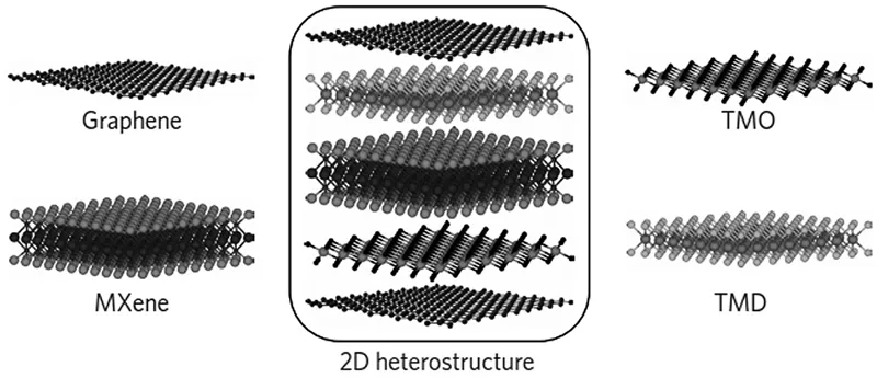
Engineered nanomaterials (NMs) are of significant interest in manufactured products. The application of these nano-dimension materials in electronics, pharmacy, and nanomedicines has an associated significant concern over their environmental impact (Erol et al., 2017; Lalwani et al., 2013). The fate and transformation of these NMs inside the human body as well as in the environment depends upon their physicochemical properties. The change or alteration in even a single set of factors such as the size, shape, or surface properties of the NM can result in a new toxicity pattern (Farré et al., 2009; Gottschalk et al., 2009; Mahmoudi et al., 2013; Podila and Brown, 2013; Powers et al., 2006). Two-dimensional nanomaterials (2D NMs) are an interesting class of NMs as the absence of the third dimension aids in exhibiting interesting attributes (Li et al., 2017; Naguib and Gogotsi, 2015). Remarkable optical and electronic characteristics have been displayed by graphene and other sister NMs such as ternary metal dichalcogenides (TMD) (Castro Neto and Novoselov, 2011; Novoselov et al., 2005). TMDs are made up of a hexagonal layer of metal atoms (M) sandwiched in between two layers of chalcogen atoms (X) (An and Yu, 2011; Coleman et al., 2011). This layered structure has strong covalent bonding within each layer (X-M-X), while a weak Van der Waals force holds the TMD sheets (MX2) (Butler et al., 2013). The exfoliation of these TMD into thin sheets provides several catalytically active surface sites for various functional applications. In the case of 2D NMs, the mobility of the charge carriers is restricted along the thickness while being permitted to transfer along the plane. The large planar area makes these materials extremely sensitive to external stimuli. Figure 1.1 exhibits the most commonly reported 2D NMs (Khan et al., 2017).
FIGURE 1.1 Schematic illustration of different kinds of 2D NMs such as graphene, transition metal carbides, nitrides (MXenes), transition metal oxides (TMO), and transition metal dichalcogenides (TMD). (From Pomerantseva, E. and Gogotsi, Y., Nature Energy, 2, 17089, 2017.)
The evolution of entirely new 2-dimensional materials with excellent features also increases the aspect of uncertainty and the necessity of understanding the toxicity profiles of these NMs. There exist multiple reports and reviews detailing various types of assays and studies on the toxicity of NMs, yet there still exists a definite deficiency in bringing them under a common domain (Chng and Pumera, 2015; Ganguly et al., 2018). Moreover, reviews summarising the cytotoxicity studies of 2D NMs are not yet reported. Standard parameters and guidelines to underscore the toxicity profile of NMs in one common domain are absent. Toxicity assaying is not a new topic but the use of NMs at an ever-increasing scale has led to questions being raised on whether the present research capacity or standard could cope in finding an appropriate answer to the toxicity profiles of such new materials. Keeping up with the rapidly changing manufacturing industries and their products has certainly raised the bar and posed a critical challenge for toxicology researchers. Safety standards and guidelines to handle such materials are critical; improper data on toxicity assessment can be fatal.
The present chapter details the different kinds of commonly observed types of assays for toxicity. The chapter also details the various physiological outcomes observed by NM interaction. Moreover, the parameters influencing the toxic profiles of 2D structures are described. Different cellular internalisation pathways are summarised. Additionally, a brief discussion of recent studies of cytotoxicity reports on 2D NMs is presented. Finally, a section detailing the safety guidelines is summarised.
1.2 Cell Death
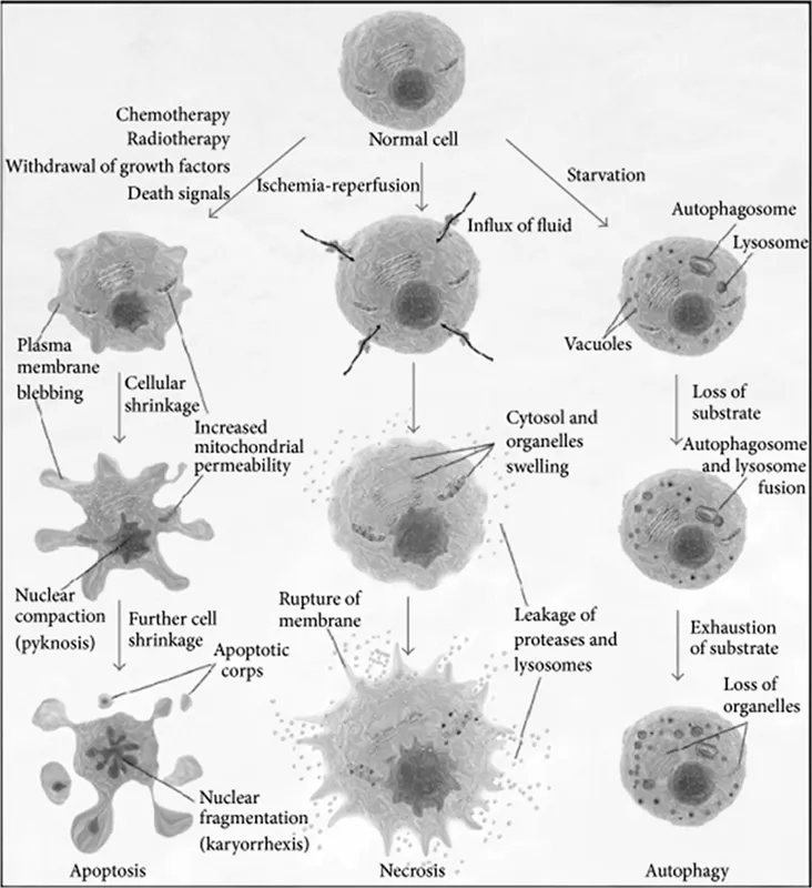
Understanding toxicity and its impact is essential for commercialising NMs. Therefore, it is vital to understand the mechanism behind cell death. The fatal mechanism of the cell is basically chartered into three types: apoptosis, autophagy, and necrosis (Figure 1.2) (Kroemer et al., 2009; Nunes et al., 2014).
1.2.1 Apoptosis
Apoptosis is commonly referred to as the type-1 cell death and relies on several cell surviving signals. The distinct property of this cell death is the complete disintegration of the cellular morphology, development of apoptotic bodies, chromatic condensation, etc. The sudden stop of these cell surviving signals triggers the apoptosis process. Intracellular proteases known as caspases are initiated once the cell surviving signals cease to exist. There exist 14 types of caspases inside a mammalian body. These caspases together initiate the cell death process (Enari et al., 1998; Galluzzi et al., 2008; Garrido and Kroemer, 2004; Kerr et al., 1972; Roach and Clarke, 2000).
FIGURE 1.2 Schematic illustration defining various types of cell death. The apoptosis pathway is represented with the characteristic cellular shrinkage and formation of the apoptotic bodies without leakage of contents. In the middle, the necrotic pathway shows the cytosol and organelle swelling and rupture of the plasma membrane with subsequent leakage of cellular contents. On the right, autophagy is illustrated with the appearance of vacuoles, the autophagosome, and its fusion with the lysosome, which ends in organelle digestion. (From Nunes, T. et al., BioMed Res. Int., 2014.)
1.2.2 Autophagy
Autophagy is called the type-2 cell death. The formation of a double-layered membrane containing vacuoles called autophagosomes initiates the type-2 cell death mechanism. The double-layered membrane engulfs the cytoplasmic components and later on combines with lysosomes to form autophagolysosomes to cause degradation (Mizushima, 2005; Tasdemir et al., 2008). This process is an intracellular degradation activity which has several pathophysiological important functions, such as tumour separation, starvation adaptation, organelle clearance, and antigen presentation. Various forms of secondary stress arise inside the cell during cell differentiation, such as nutrient starvation, which initiates the process of autophagy (Baehrecke, 2002; González-Polo et al., 2005; Levine and Kroemer, 2008; Mizushima, 2007).
1.2.3 Necrosis
Necrosis is the type-3 cell death process known as uncontrolled cell death process. It arises due to physicochemical stress initiated by an alteration in the nucleus—for example, karyolysis, pyknosis, etc.—and in the cytoplasm (condensation, rupture of cytosol, and further disintegration). Necrosis is the process that arises 12–20 hours after the cell death; hence, it cannot be necessarily defined as cell death. However, the other two types of cell death initiate the process of necrosis. Necrosis results in cytoplasmic swelling, dilation of organelles, and rupture of the plasma membrane, which results in the outflow of cytosolic content and damage of the outside cellular environment (Festjens et al., 2006; Golstein and Kroemer, 2007; Majno and Joris, 1995).
1.3 Different Types of Assay
1.3.1 In Vitro Assay
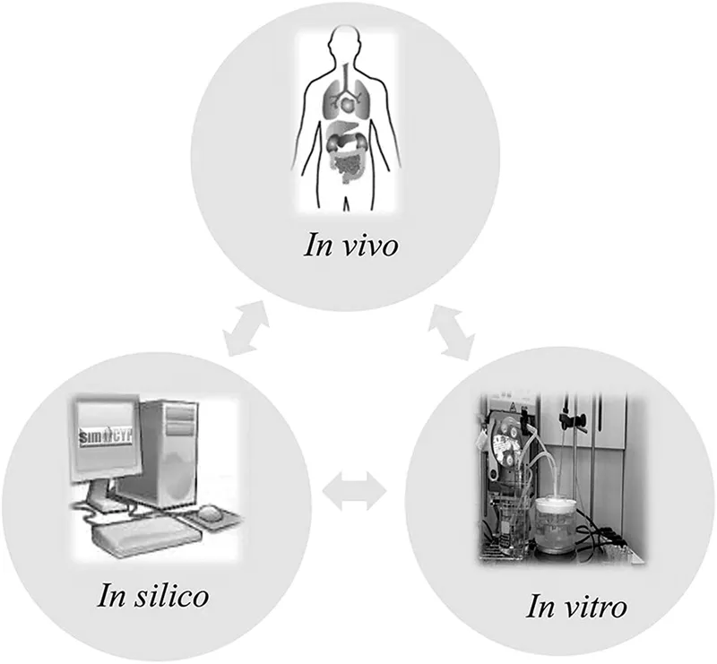
One of the major toxicity assessment techniques, in vitro assay is used to evaluate the toxicity of materials (Figure 1.3) (Beloica et al., 2015). Different cell lines are exposed against several xenobiotic agents and incubated for definite time intervals. This potentially helps in calculating the dosage of the exposure. Different assays are used to measure the proliferation and cellular metabolism such as 3-(4,5-Dimethyl-2-thiazolyl)-2,5-diphenyl-2H tetrazolium bromide (MTT) and 2-(2-methoxy-4-nitrophenyl)-3-(4-nitrophenyl)-5-(2,4-disulfophenyl)-2H-tetrazolium, or mono sodium (WST-8). These techniques are rapid and cost-effective and do not require the use of animals, which reduces any form of ethical conflicts. However, these processes are supposed to mimic the cellular environment, which does not always correlate to the physiological outcomes (Ciappellano et al., 2016; Lewinski et al., 2008; Roggen, 2011; Sharifi et al., 2012; Stockert et al., 2012; Wataha et al., 1991).
FIGURE 1.3 Illustration defining different types of toxicity assays. (From Beloica, S. et al., Eur. J. Pharm. Sci., 75, 151–159, 2015.)
1.3.2 In Vivo Assay
This toxicity assessment technique is considered the most reliable method to date (Figure 1.3). A small amount/dose of the xenobiotic component is administered in model animals such as mice. The component’s transportation, metabolism, distribution, and finally removal are the key aspects for the concern. This method requires monetary as well as time investments. However, the results obtained are highly efficient and reliable. Concerns of ethical conflicts persist with this kind of preclinical trial (Aillon et al., 2009; Fielden and Kolaja, 2008; Filip et al., 2015; Lee et al., 2016; Wen et al., 2017).
1.3.3 In Silico Assay
Utilising several theoretical models to judge the physicochemical properties of some xenobiotic materials is one of the novel routes of assaying (Figure 1.3) (Hindman and Ma, 2018). Formation of quantitative structures to collectively develop toxicity assaying models is one of the key elements. These quantitative structures are gathered from existing literature and mathematical modelling studies. This method of toxicity assaying is cost effective, less time consuming, and free from any possible ethical conflicts. A wide gap exists in predicting the behavioural pattern using theoretical modelling based upon the properties of this ever-changing material. Additional experimental validation using one of the other two assaying techniques is essential (Aires et al., 2017; Bell et al., 2018; Gao et al., 2018; George et al., 2011; Gleeson et al., 2012; Laomettachit et al., 2017; Sizochenko et al., 2018).
1.4 Physiological Impacts Due to Nanomaterials Interaction
1.4.1 Oxidative Stress and ROS Generation
The ROS levels in the human body are critical for different metabolic functions (Jornot et al., 1998; Sharifi et al., 2012). Variations in the ROS levels can cause alteration of key cellular events such as signal transduction and protein redox potential. ROS are generated when the cellular redox potential is comparable to the band edge potentials of the interacting nanoparticles (Choi, 2016; Nel et al., 2006; Stahl et al., 1998). The neutralising mechanism is initiated inside the cell, which is defined as oxidative stress. Redox signalling pathways like mitogen-activated protein kinase (MAPK) cascades are enabled in this condition (Jornot et al., 1998). Several cellular processes such as cell proliferation and differentiation are controlled by these protein cascades (Apel and Hirt, 2004). Proinflammatory cytokines and chemokines are expressed with the help of these protein cascades. At tier 1, lower levels of oxidation stress aid in the expression of genetic antioxidant response, which results in the formation of antioxidant enzymes. These enzymes are responsible for the activation of the transcription factor Nrf2 (Li et al., 2004; Sun et al., 2017). These transcription factors are responsible for tier 2 stress mitigation by expressing many anti-...