Thanks to tremendous technical advances in molecular biology and cellular imaging after those in electrophysiology, there is now a deep understanding of the physiology of nerve cells and their synaptic interconnections. The complexity of the brain emerges from the communication and interaction between billions of these elements. This book explores systematically and didactically the details of neuronal physiology, covering membrane biophysics, receptor physiology, sensory transduction and synaptic transmission with its selective pharmacology. Readers of the book will be fully equipped to understand the functions and possibilities of the key units of the brain's parallel computations.
- 458 pages
- English
- ePUB (mobile friendly)
- Available on iOS & Android
eBook - ePub
Physiology of Neurons
About this book
Trusted by 375,005 students
Access to over 1.5 million titles for a fair monthly price.
Study more efficiently using our study tools.
Information
CHAPTER
1
Signaling in the Brain: An Introductory Chapter
Why a textbook specifically concerned with neurobiology as a complement to a textbook on cell physiology? What are the distinct aspects of the neural tissue that one has to comprehend to understand its originality? This introductory chapter briefly presents the characteristics of the neuronal tissue on which we will focus, therefore outlining the framework of this book.
Each single neuron is polarized: in its apical dendritic region, either it processes a physical signal decoding its parameters or it integrates a series of input signals arising from other neuronal structures. The emerging signal is then vehiculated over a variable distance depending on the neuron morphology toward its axonal ending, and it is there delivered locally at synapses, contact points with other neurons. The information carried all along a neuron is an electrical signal, graded in the dendrite and all or none in the axon where action potentials propagate, carrying the efferent message in a binary way. Only at the synaptic endings are the message characteristics delivered by release of a chemical neurotransmitter that in turn will excite or inhibit locally a postsynaptic neuron. Already at this point, the notion of multiple connections appears. Between the periphery, where we contact the outside world to the brain, and the brain, where our perception is elaborated and decisions taken to correlate events and/or deliver a movement, a number of connected relay neurons are involved. When using the panoply of stainings presently at our disposal to reveal these connections, it appears that the neuronal network is built on such series of neurons. The simplifying grouping of similar nerve cells in layers favors an additional one in parallel connectivity. To this relatively simple organization is further added a number of loops, forward and backward, whose effects will slightly vary from one cell to the other, and from one activation of the system to the next with new properties possibly arising upon multiple iterations. Despite similarities between some neuronal cells, the emerging picture is that of a highly complex nervous system.
The following chapters will present the state of the art of our present understanding of the signaling properties of the neuronal cells, the building blocks of the brain. Starting at the single-cell level, one already has a good grasp on the transduction processes occurring in the sensory neurons; the steps leading to coordinated muscular contractions generating body movements such as walking, speaking, and breathing are precisely described. Understanding pre- and postsynaptic transmission processes clarifies the numerous effects of the neuroactive drugs and hormones. Progressively the reader will acquire the complex terminology of neurobiology that pertains to so many domains: anatomy, biophysics, molecular biology, etc. This book aims to be a guide to understand from the bottom up how neurons and the brain work. Its reader should be able to take advantage of recently developed tools to devise experiments allowing us to fill the gap between what we know to occur at the cellular level and the functioning of neuron networks.
If the exquisitely complex properties of the neuronal network can only be reached by the modeling approach, which is rightly taking on more and more importance in neurobiology, we are convinced that performant models can only be developed by knowing as accurately as possible the details of the properties of individual neurons as presented in this book. Its reader will be equipped with the quantitative biophysical knowledge that is a prerequisite to realistic computation studies of neuronal and network function.
1.1 Neurons and Glial Cells Build Up the Nervous System
In vertebrates, the nervous system (NS) is divided into a central nervous system (CNS), the brain in the head and the spinal cord, and the peripheral nervous system (PNS), which is made up of the nerves connecting the CNS to the peripheral organs, muscles, and all viscera. The anatomy of the brain is complex. Neuroanatomists divide it into six main regions: the telencephalon (cerebral hemispheres), diencephalon (thalamus and hypothalamus), mesencephalon (midbrain), cerebellum, pons, and medulla oblongata. A simplified presentation of the brain is given in Annex A with brain sections oriented following the conventions of histology. The stainings of these brain slices show structures or brain nuclei mainly constituted by cell bodies and some cellular extensions, with tracts of fibers impinging on them or extending out of them. This coarse organization appears when using a basophilic staining with methylen blue, which reveals the abundant ribosomes accumulated around the cell nucleus where an important biosynthetic activity takes place. The nuclei constitute the gray matter, named after the gray color of the numerous cell bodies they contain. Slices show regions of white matter, made of the bundles of myelinated fibers connecting NS nuclei to distant targets: these axons have a sheath containing myelin, a lipid-rich substance with insulating properties, yielding a pearl white color. A determinant step in our understanding of the functioning of the NS was the introduction of individual cell staining in the 1870s by Camillo Golgi. In the Golgi staining, silver nitrate and potassium dichromate are added to a tissue slice, eventually provoking an initial metallic precipitate of silver chromate. Interestingly, precipitation may extend over all the ramifications of the cell on which it has started. Using this staining systematically on different brain slices, Santiago Ramon y Cajal described neuronal entities with distinct complex morphologies. He finally proposed the cell theory (as opposed to the reticular theory), which describes neurons as non-interconnected cells, constituting independent units of the nervous system. With Golgi in 1906, Ramon y Cajal was awarded the first Nobel Prize attributed to neurobiologists.
Since then, many staining techniques have been developed that have helped to characterize individual cell morphology. Cells with their ramifications and projections can be loaded by intracellular microinjections of a fluorescent dye or of biocytine. A live cell labeled with a fluorescent dye can be visualized with the use of adapted optics. Contours of a biocytin labeled cell will be studied after fixation. Cells can be labeled also extracellularly: fluorescent carbocyanines, for instance, dissolve in the lipophilic membrane of a cell and diffuse over its whole surface, thus revealing its contours (Figure 1.1b). These techniques have proved valuable for the localization of a neuron previously characterized electrophysiologically and for the identification of cell projections.
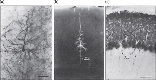
A number of other techniques also aim at identifying cell function. In situ hybridization has been extensively used to characterize neurons by tagging the mRNA coding for a specific receptor or channel. Antibodies directed against a specific cell component, either intracellular or in the membrane, will label the entire neuron. In Figure 1.1c, the morphology of the dendritic tree of a Purkinje cell is revealed by an antibody directed against calbindin, a Ca2+-binding protein abundant specifically in this cell type.
These stainings have revealed the diversity of morphologies and hodologies of the cells in NS structures. As we will see, two main types of cells operate in the brain: in addition to neurons, glial cells are the other actors operating in the NS.
1.1.1 A neuron is a cell equipped with dendrites and an axon
A neuron, such as the pyramidal cell of the cerebral cortex (Figure 1.1) has features common to all neurons throughout the whole nervous system. The cell body (or soma) contains the nucleus and the ribosomal machinery. Out of it extend two types of processes: from one pole of the cell body emerges a thin process, the axon, which can be very long depending on the target cell it is connected with. More or less directly from the other pole of the cell body emerge branched, bushy ramifications, the dendrites: they receive inputs from other brain regions. This organization varies slightly from one cell type to another with the dendrites mainly being differentially distributed. Dendrites can emerge from all over the soma, either forming a single bush grown on a main process or being distributed in a few bushes. In the Purkinje cell of the cerebellum, the synaptic contacts are localized on a large dendritic tree (Figure 1.1c). As a noteworthy exception, let us cite the somatosensory neurons: their soma is located in the dorsal root ganglia. A single process extends out of this soma, which very closely bifurcates: an axonal branch projects to the spinal cord, whereas the other branch goes toward the periphery. In contrast, the axon is absent in photoreceptors. In invertebrates, often a single process comes out of the cell body and the synaptic contacts occur on proximal branches, the axon then running as a prolonged thin extension. Despite these exceptions, the prototypical neuron is described as a cell carrying two distinct processes emerging from its soma: dendrites at one end and an axon at the other end.
Ramon y Cajal observed many cell types, including sensory cells, and concluded that the neurons were probably functionally polarized, the information going from the receivers on the dendrite to the axon ending. As we will see, this feature has been largely confirmed since. The area of contact of two neurons is called a synapse. Taking into account the polarization of the information, one neuron is presynaptic to the other one, which is postsynaptic to it. In most cases, the message carried by a neuron is delivered at its end by synaptic machinery, which releases a neurotransmitter, a chemical signal that affects the activity of the juxtaposed dendrite: facing the synaptic terminal, spines, small protrusions of the postsynaptic dendrites, contain receptors sensitive to the released transmitter. These characteristics are schematically summarized in Figure 1.2.
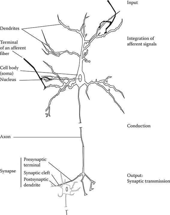
Table of contents
- Cover
- Half Title
- Title Page
- Copyright Page
- Contents
- Detailed Contents
- Foreword
- Preface
- Contributors
- Chapter 1: Signaling in the Brain: An Introductory Chapter
- PART I: ION MOVEMENT IN THE CELL
- PART II: FAST AND SLOW NEURONAL SIGNALING
- PART III: SYNAPTIC TRANSMISSION
- PART IV: METHODS
- Index
Frequently asked questions
Yes, you can cancel anytime from the Subscription tab in your account settings on the Perlego website. Your subscription will stay active until the end of your current billing period. Learn how to cancel your subscription
No, books cannot be downloaded as external files, such as PDFs, for use outside of Perlego. However, you can download books within the Perlego app for offline reading on mobile or tablet. Learn how to download books offline
Perlego offers two plans: Essential and Complete
- Essential is ideal for learners and professionals who enjoy exploring a wide range of subjects. Access the Essential Library with 800,000+ trusted titles and best-sellers across business, personal growth, and the humanities. Includes unlimited reading time and Standard Read Aloud voice.
- Complete: Perfect for advanced learners and researchers needing full, unrestricted access. Unlock 1.5M+ books across hundreds of subjects, including academic and specialized titles. The Complete Plan also includes advanced features like Premium Read Aloud and Research Assistant.
We are an online textbook subscription service, where you can get access to an entire online library for less than the price of a single book per month. With over 1.5 million books across 990+ topics, we’ve got you covered! Learn about our mission
Look out for the read-aloud symbol on your next book to see if you can listen to it. The read-aloud tool reads text aloud for you, highlighting the text as it is being read. You can pause it, speed it up and slow it down. Learn more about Read Aloud
Yes! You can use the Perlego app on both iOS and Android devices to read anytime, anywhere — even offline. Perfect for commutes or when you’re on the go.
Please note we cannot support devices running on iOS 13 and Android 7 or earlier. Learn more about using the app
Please note we cannot support devices running on iOS 13 and Android 7 or earlier. Learn more about using the app
Yes, you can access Physiology of Neurons by Anne Feltz in PDF and/or ePUB format, as well as other popular books in Biological Sciences & Diseases & Allergies. We have over 1.5 million books available in our catalogue for you to explore.