A Practical Approach to Medical Image Processing
eBook - ePub

A Practical Approach to Medical Image Processing

Elizabeth Berry

  1. 304 pages
  2. English
  3. ePUB (mobile friendly)
  4. Available on iOS & Android
eBook - ePub

A Practical Approach to Medical Image Processing

Elizabeth Berry

Book details
Book preview
Table of contents
Citations

About This Book

The ability to manipulate and analyze pictorial information to improve medical diagnosis, monitoring, and therapy via imaging is a valuable tool that every professional working in radiography, medical imaging, and medical physics should utilize. However, previous texts on the subject have only approached the subject from a programming or computer science viewpoint at a mathematically inaccessible level. Unlike these previous publications, A Practical Approach to Medical Imaging Processing provides hands-on instruction, using the freely available software program ImageJ, on all of the skills needed to perform filtering and image enhancement techniques used in structured image discrimination.

In this unique text, the author focuses exclusively on image processing and treats medical images in a generic way to highlight the features that all digital images have in common. The book first introduces the main topics in image processing and as it progresses, you will discover relevant points of good practice. The author validates each technique with a corresponding case study, which originates from a published journal article. The case studies demonstrate how the concepts of image processing are applied to real-life situations, such as how to uncover information suffering from distortion and pixel-size limitations. The accompanying downloadable resources contain the Windows version of the ImageJ software, digital images, and documents to be used during the practical activities included in each chapter.

With its highly functional workbook approach, A Practical Approach to Medical Image Processing allows you to build your skills in image manipulation and to enjoy the benefits of this valuable field without having to code or develop your own program.

Frequently asked questions

How do I cancel my subscription?
Simply head over to the account section in settings and click on “Cancel Subscription” - it’s as simple as that. After you cancel, your membership will stay active for the remainder of the time you’ve paid for. Learn more here.
Can/how do I download books?
At the moment all of our mobile-responsive ePub books are available to download via the app. Most of our PDFs are also available to download and we're working on making the final remaining ones downloadable now. Learn more here.
What is the difference between the pricing plans?
Both plans give you full access to the library and all of Perlego’s features. The only differences are the price and subscription period: With the annual plan you’ll save around 30% compared to 12 months on the monthly plan.
What is Perlego?
We are an online textbook subscription service, where you can get access to an entire online library for less than the price of a single book per month. With over 1 million books across 1000+ topics, we’ve got you covered! Learn more here.
Do you support text-to-speech?
Look out for the read-aloud symbol on your next book to see if you can listen to it. The read-aloud tool reads text aloud for you, highlighting the text as it is being read. You can pause it, speed it up and slow it down. Learn more here.
Is A Practical Approach to Medical Image Processing an online PDF/ePUB?
Yes, you can access A Practical Approach to Medical Image Processing by Elizabeth Berry in PDF and/or ePUB format, as well as other popular books in Physical Sciences & Physics. We have over one million books available in our catalogue for you to explore.

Information

Publisher
CRC Press
Year
2007
ISBN
9781000738315
Edition
1

1

Image Processing Basics

1.1 Introduction

In this introductory chapter the basic terminology of image processing is presented. The ImageJ program is introduced using practical exercises, so that the reader is able to perform image processing tasks. Fundamental imaging processing principles, covering both the grayscale and spatial properties of an image, are described. The chapter closes with an overview of the content of the book, which is based around the five classes of image processing: image enhancement, image restoration, image analysis, image compression and image synthesis.

1.1.1 Learning Objectives

When you have completed this chapter, you should be able to
  • Define the important basic terms used in image processing
  • Understand methods of image enhancement that use the histogram
  • List several different measurements, both spatial and grayscale, which can be made from images
  • Discuss the five classes of image processing
  • Use ImageJ to load and save an image, and perform simple tasks

1.1.2 Example of an Image Used in This Chapter

The first self-assessment question in each chapter is based on one of the images that appears later in the chapter. The question usually requires some background knowledge of medical imaging technology and is intended to help those who have previously studied medical imaging to start to integrate medical image processing with their existing knowledge. Those who lack this background knowledge should not be concerned if they cannot answer the first question in each chapter, and note that no prior knowledge is assumed in the other questions in each chapter.
Image
FIGURE 1.1
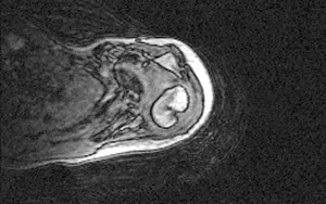
Image of a shoulder for self-assessment question 1.01.
Self-assessment question 1.01
Figure 1.1 is an example of an image used in this chapter. It shows the shoulder joint. The original image was 256 × 160 pixels in size and each pixel represented a 0.8 mm cube of tissue. Which imaging modality has been used to acquire the image shown? Note the loss of image information towards the left of the image. What does this suggest about the particular equipment that was used to acquire the image?

1.2 Definition of Image Processing

Image processing refers to the manipulation and analysis of pictorial information, for example by improving, correcting, changing or analyzing the image. The simplest example is the action of prescription spectacle lenses, which are designed to counteract distortion caused by defects in the owner’s eye. The action of the lens is called an optical process because the lens is acting on an optical image. Optical images can be converted to analog images. An analog image is a two-dimensional representation of a scene; the image contains continuously varying tone (e.g., in a monochrome or black and white photograph) or continuously varying color.

1.2.1 The Digital Image

An analog image can be sampled to give an array of discrete points, each of which has an associated brightness value. This is a digital image, which is simply an array of numbers, one for each sample point (Figure 1.2). Each sample point is known as a picture element, or pixel. An image is generally sampled using a rectangular array of pixels. The process of sampling and assigning the numerical value is called digitization. Thus a digital image is an array of pixels, each of which has a value. When these data are displayed in the correct arrangement, with each pixel value used to give the brightness or color at that point, we see the image. The image size, or matrix, is expressed in terms of the number of pixels in the rectangular array, for example 256 × 256 pixels. When image processing is performed on such an image, it is more fully described as digital image processing. In medicine, some digital images are acquired in digital form from the outset (e.g., computed tomography), while others are acquired as analog images (e.g., a chest x-ray on film) that may later be digitized. Three-dimensional data sets consist of a series of images each representing a slice through the object. In this case pixels are known as voxels.
Image
FIGURE 1.2
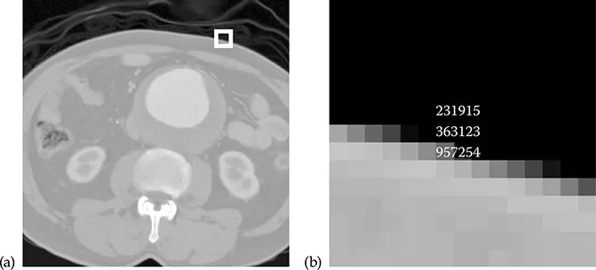
A digital image is an array of pixels, each of which has a value. (a) A digital image showing the location of a small square region of 15 × 15 pixels. (b) An enlargement of the 15 × 15 region of the image. The numerical values associated with the central pixels are shown overlaid.

1.2.2 Image Resolution

The resolution of an image is a measure of the fidelity (faithfulness) of the representation of the original scene. Resolution is related firstly to the characteristics of the imaging system and secondly to the number of pixels and to the range of brightness values that are used for digitization. In the discussion in this chapter, it will be assumed that the digitization process is performed on a perfect representation of a scene, so the properties of the imaging system may be ignored. The effects of the imaging system are considered in Chapter 7.
The question addressed when considering image resolution is “Does the digital image resolve elements of the original scene?” There are two components of resolution: grayscale resolution and spatial resolution. The question therefore has two parts: Can objects that are similar in tone or color be distinguished? Can small objects be seen?
1.2.2.1 Grayscale Resolution
The digitization process assigns a number to each pixel to represent the brightness at that location in the original image. The grayscale, or brightness, resolution is affected by how many brightness values are allowed. It is very common to assign the pixel value to one of 256 discrete values (from 0 to 255). Each value is displayed as a separate shade of gray, and these shades run smoothly from dark (0) to light (255). An image with 256 discrete values is known as an 8-bit image, because 28 = 256. An 8-bit grayscale is shown in Figure 1.3a.
Image
FIGURE 1.3
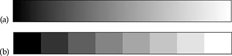
Grayscales associated with images of two different bit depths. (a) An 8-bit grayscale (256 ...

Table of contents

  1. Cover
  2. Half Title
  3. Series Page
  4. Title Page
  5. Copyright Page
  6. Dedication
  7. Table of Contents
  8. Supplementary Resources Disclaimer
  9. Preface
  10. Acknowledgments
  11. On the CD
  12. 1 Image Processing Basics
  13. 2 Segmentation and Classification
  14. 3 Spatial Domain Filtering
  15. 4 Frequency Domain Filtering
  16. 5 Image Analysis Operations
  17. 6 Image Data Formats and Image Compression
  18. 7 Image Restoration
  19. 8 Image Registration
  20. 9 Visualization and 3-D Methods
  21. 10 Good Practice
  22. 11 Case Studies
  23. 12 For Instructors
  24. Index
Citation styles for A Practical Approach to Medical Image Processing

APA 6 Citation

Berry, E. (2007). A Practical Approach to Medical Image Processing (1st ed.). CRC Press. Retrieved from https://www.perlego.com/book/1507459/a-practical-approach-to-medical-image-processing-pdf (Original work published 2007)

Chicago Citation

Berry, Elizabeth. (2007) 2007. A Practical Approach to Medical Image Processing. 1st ed. CRC Press. https://www.perlego.com/book/1507459/a-practical-approach-to-medical-image-processing-pdf.

Harvard Citation

Berry, E. (2007) A Practical Approach to Medical Image Processing. 1st edn. CRC Press. Available at: https://www.perlego.com/book/1507459/a-practical-approach-to-medical-image-processing-pdf (Accessed: 14 October 2022).

MLA 7 Citation

Berry, Elizabeth. A Practical Approach to Medical Image Processing. 1st ed. CRC Press, 2007. Web. 14 Oct. 2022.