![]()
CHAPTER 1
Introduction
O ptical microscopes are probably the most widely used instrumentation across all branches of science and medicine, and play a significant role in helping to advance human understanding through research as well as being an everyday tool for quality control in industry and diagnosis in the clinic. They perhaps provide the ultimate implementation of the idiom “seeing is believing” as microscopy has performed a crucial part in helping us explore and understand a world where structures and organisms are smaller than can be seen directly with the human eye. For hundreds of years optical microscopes and telescopes have paved the way for new scientific insights at both ends of the length scale. The desire for ever higher quality images and improved resolution has led to significant advances in both theoretical optical physics and practical engineering and, as new photonics technology has been invented, this has rapidly been applied in optical microscopy. In the last twenty years there has been a significant upsurge in developments in the field of optical microscopy leading to a vast array of new imaging modalities, each of which is excellent for one imaging task but may be less suitable for another. The aim of this book is to provide a simple guide in helping to select the most suitable method of optical microscopy to use for a particular application and the subsequent use of the preferred instrumentation.
To make the book as useful as possible to a wide range of readers each chapter is self-contained with the physical concepts behind a specific imaging process being explained through marked areas of text. In order to appreciate how a method is best applied to a particular task it is not necessary to read these more detailed physical insights. Although the text is generally focused on the application of microscopy to biological samples the general principles described are broadly applicable and where possible suitable examples from geology and materials science are given. Although huge technological advances in optical microscopy methods have been made in the last twenty years the core “physics of imaging” has not changed and the limitations that these play in microscopy will be highlighted throughout. An appreciation of these principles is very helpful to understand the trade-offs that always have to be made between spatial resolution, speed of imaging and the level of perturbation to the sample in order to obtain the images required. The underlying ethos used throughout is that of selecting the correct tool for the particular task; if results can be achieved with a low-cost camera and magnifying lens then this is the route to select. The highly complex and technical methods can then be applied to the tasks that really require that level of sophistication. However, before presenting the details of both the imaging process and subsequent practical implementation of the methods it is worth considering optical microscopy in a historical perspective.
1.1 A Historical Perspective
Being able to observe events has always been the cornerstone of scientific discovery ranging from monitoring the movement of the sun, moon and stars through to watching the tiniest visible creatures navigate their way around the world. This desire to see events at different scales has, throughout the centuries, advanced optical instrumentation and, as will be shown shortly, provided a significant boost to basic understanding of light and optics. Such visual observations were initially undertaken using just the human eye. Since the invention of the lens around 2000 years ago, there has been a slow refinement in the ability to image with ever greater detail at both astronomical and miniature scales. Perhaps the first recorded use of the lens for making observations at a microscopic scale was by Pliny the Elder (in Bostock, 1855) where a magnifying lens was used to look at tissue damage at a fine scale and also, in some cases, to cauterize the wounds using sunlight. At this stage clearly the optical physics taking place to produce the magnified image was not understood and the lenses were produced through the controlled melting of glass to form droplets, with luck as well as skill being involved in the final quality of the product. Although observations on the effect of beads of glass, and indeed water filled containers, were made by scholars such as Ibn al-Haytham and Roger Bacon, practical applications of the magnifying effect were not really considered, or at least recorded, until Salvino D’Armate in Italy during the 12th century. Using a single positive lens, which he was making to help correct the loss of close sight with age (presbyopia), he noted that the world could be enlarged to the viewer. There is however little further information on what was observed in this new “microscopic” world.
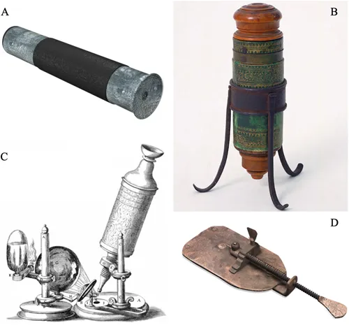
Around 300 years later, in 1590, when the craft of spectacle making was firmly established, Zacharias Jansen and his father Hans, in the Netherlands, created what is generally viewed as being the first optical microscope. This was a compound instrument consisting of multiple lenses developed entirely through experimentation and carefully recorded observations. Although all their original notes were lost during the Second World War it is known they developed an instrument that consisted of three tubes which were moved for focusing and altering the magnification (Figure 1.1A). The eyepiece was a bi-convex lens and the objective a plano-convex lens, which required the most advanced polishing methods available in the late 16th century. Perhaps this is the first example of the microscope making use of an optical technological advance. Based upon the notes made the magnification could be adjusted to be between three and ten times, depending on the position of the sliding tubes and working distance. Although crude by modern standards, both in terms of optical design and fabrication, it opened up the world of the very small for the first time. It is known that Galileo Galilei also was experimenting with compound optical systems to develop his first telescope and was aware of the advances being made in the Netherlands. In 1609, he developed a compound microscope using both convex and concave lenses. He described his “modified telescope to see objects very close” in his book The Assayer (Il Saggiatore) (1623) and several variations of this instrument were built (Figure 1.1B). It was around this time that the term “microscope” was first used.
FIGURE 1.1 A) One of the original Jansen microscopes, B) Galileo Galilei’s original microscope, C) Drawing of Hooke’s microscope; note the oil lamp and large lens for improved illumination, D) A copy of Anton van Leeuwenhoek’s single lens, ×200 magnification microscope. (Images A and D credit to Molecular Expressions.com at Florida State University, Image B Credit Museo Galileo).
Now that the basic compound microscope had been developed, for the next few hundred years it was the application of this new instrument that became increasingly important as people started to look at a wide range of samples. Robert Hooke, while the Curator of Experiments of the Royal Society, published his findings (1665) in which the term “cell” was used for the first time to describe what he thought of as the basic unit of life. The images contained in his book, Micrographia, were taken with one of the best microscopes of the time and the illustration of its operation (Figure 1.1C) demonstrates the importance of the illumination system occupying almost as much of the image as the actual observation optics. It is perhaps interesting to note here that for many of the most advanced systems in use today described in the following chapters the light source still occupies a major physical area in the laboratory! Although best known for the high quality copperplate engravings (from Hooke’s original drawings) the book also describes the wave theory of light, the organic nature of fossils and astronomical observations. Some of these writings caused him to come into conflict with Isaac Newton and it is only in the 20th century that perhaps the overall scientific importance of Robert Hooke has been appreciated.
Improvements to the microscope were then made through advances in lens fabrication led by Anton van Leeuwenhoek in the Netherlands. His microscope consisted of a single ball lens and the sample was mounted on a pin that could be finely adjusted to bring the sample into focus (Figure 1.1D). Due to his ability to produce high quality lenses, through carefully controlled melting and pulling of glass, he produced systems with magnification up to around 250 times. By comparison compound microscopes were limited to around 30 times magnifications before distortions, caused by defects in the glass used in larger lenses, led to blurred images. Van Leeuwenhoek undertook observations on multiple live samples and is credited with discovering single cell organisms or “animalcules”. At the time his findings were treated with scepticism, in particular by the Royal Society, though they did eventually publish his observations (around 560 letters) and made him a Fellow. This only occurred after a team from London went out to the Netherlands to observe his microscopes in operation to ensure he was not “inventing” this new microscopic world!
Slowly over the next 200 years the ability to produce high quality optical elements improved, along with a basic understanding of how lenses should be brought together for improved image quality. In particular in 1826 Joseph Lister developed achromatic doublet lenses removing one of the major observable aberrations present in these systems. However, even then most of the designs were based upon experimentation and trial and error.
Advances in the understanding of optics were being made with greater understanding of the mathematics behind light propagation and the behavior of waves. However, the whole field of optics was then revolutionized through trying to build better microscopes. Ernst Abbe working for Carl Zeiss in Jena developed the framework for the full performance, and limitation, of optical imaging instruments (Abbe 1873) and later presented his findings to the Royal Microscopical Society (Abbe 1881). Abbe’s findings were largely based upon careful experimentation while around the same time Herman Helmholtz was working on a theoretical approach. Joseph Lagrange, more than 60 years previously, had hinted at the limitations through mathematically based reasoning. As a result of this discovery Abbe became a shareholder in the Zeiss company becoming very wealthy, though he did also introduce the eight-hour day into the Zeiss workshop. He proved that the maximum resolution possible is proportional to the wavelength and inversely proportional to the numerical aperture (light gathering power) of the lens. Details on the physical aspects of this fundamental limitation are provided in Chapter 2. Abbe’s work at this time advanced the entire optical field for practical devices and instruments and although huge technical innovations have been made in optical microscopes since this date the majority of improvements have been through technology and engineering rather than fundamental changes in optical design.
In 1902 Richard Zsigmondy was interested in the study of colloidal gold suspensions and wanted to observe the movement and position of particles that were smaller than Abbe’s limit of resolution. Working with Henry Siedentopf (an employee of Zeiss) he developed a microscope in which a sheet of light illuminated the sample and the observation of the light scattered by the particles was observed orthogonally to the excitation. This technique of “ultramicroscopy” (Siedentopf and Zsigmondy 1902) was a method really ahead of its time as the observations all had to be made by eye since photography was not sufficiently advanced and CCD cameras not even a dream. However, the results of Zsigmondy’s observations were sufficient for him to be awarded the Nobel Prize for Chemistry in 1925. The method was rediscovered as light sheet or selective plane illumination microscopy (Huisken et al. 2004). This preferred method of imaging in vivo dynamic events especially for extended periods of time is described in Chapter 7.
Subsequent developments in basic optical microscopy have generally been in methods of improving the contrast in the image rather than significant changes in optical design. In this context the next major advance was in the development of phase contrast microscopy by Fritz von Zernike in 1934 (Zernike 1934) which is described in detail in Chapter 4. Zernike was awarded the 1953 Nobel Prize for Physics for this breakthrough and he acknowledged the role that Abbe’s work had played in his discovery. As other methods of seeing minute structures developed, such as electron microscopy, with resolution that was better than that offered by optical methods, people started to consider what the real advantages were in optical microscopy. This should still be the first question asked when considering how to observe a very small structure or process.
A major benefit of the optical microscope is clearly the ease of use, at least for basic systems, alongside the simple...