- 474 pages
- English
- ePUB (mobile friendly)
- Available on iOS & Android
eBook - ePub
About this book
This is a vital revision aid for postgraduate radiology students taking the Fellowship of the Royal College of Radiology (FRCR) Part 2 final exams. Part 2 comprises two elements: 2a includes a series of six multiple choice exams covering the major body systems, and 2b contains a written exam and an oral viva typically taken at the beginning of the fourth year of specialty training.
Tools to learn more effectively

Saving Books

Keyword Search

Annotating Text

Listen to it instead
Information
chapter 1
Cardiothoracic and Vascular
Cardiovascular
Aberrant left pulmonary artery
Occurs due to the failure of formation of the sixth aortic arch. Blood to the left lung arises from an aberrant left pulmonary artery that arises from the right pulmonary artery. The vessel passes between the trachea and oesophagus and causes narrowing of the trachea in a caudal direction. Associated with other anomalies (e.g. patent ductus arteriosus).
Plain film
●Bronchial obstruction causes lung emphysema (right lung, middle lobe, lower lobes, left upper lobe)
Barium swallow
●Anterior indentation on the oesophagus, just above the level of the carina
Aortic aneurysm
Considered either true (aneurysm bound by all three walls of the vessel) or false (i.e. pseudoaneurysm, part of the wall of the aneurysm is formed by surrounding soft tissue). Aneurysms are described as being saccular or fusiform.
●Saccular aneurysms are eccentric in shape, the aneurysm only forming from part of the circumference of the vessel wall. Associated with mycotic aneurysms (Figure 1.1).
●Fusiform aneurysms involve the full vessel circumference and feature cylindrical dilatation. More commonly seen with atherosclerotic aneurysms (Figure 1.2).
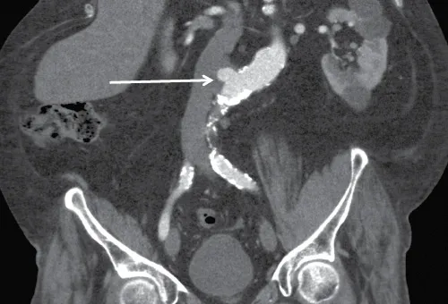
Figure 1.1 Saccular aneurysm. CT angiogram demonstrating a saccular aneurysm arising from the abdominal aorta.
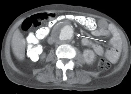
Figure 1.2 Fusiform aneurysm. CT angiogram demonstrating a fusiform abdominal aortic aneurysm.
CT
●Thoracic aortic aneurysms are mostly atherosclerotic and calcified in 75%. Other causes include cystic medial necrosis (a disorder of the large arteries with formation of cyst-like lesions in the media, associated with e.g. Marfan and Ehlers–Danlos syndromes) and syphilis (expect extensive calcification).
●Abdominal aortic aneurysms (AAAs)—mostly atherosclerotic.
●Popliteal aneurysms, associated with an AAA in 30%–50%.
Intervention
●Advised when diameter >5.5 cm (the risk of rupture is greatly increased over this).
●Endovascular stents are generally oversized by 10%. The presence of perigraft air is a common finding in the immediate post-operative period; however, if present >1 week after surgery, suspect infection.
●Endoleak is defined as the continued perfusion of the aneurysm despite placement of a stent graft (Table 1.1).
Table 1.1 The classification of endovascular stent graft endoleaks
| Type of endoleak | Site |
| Type 1 | Leak from the stent/graft attachment due to an inadequate seal |
| 1a | Proximal |
| 1b | Distal |
| Type 2 (most common, 80%) | Filling of the sac from retrograde flow through aortic branches (e.g. lumbar arteries, inferior mesenteric) |
| Type 3 | Structural failure of the stent graft/leak from mid-g... |
Table of contents
- Cover
- Half Title
- Title Page
- Copyright Page
- Contents
- Foreword
- Acknowledgements
- Introduction
- Authors
- 1. Cardiothoracic and Vascular
- 2. Musculoskeletal
- 3. Gastrointestinal
- 4. Genitourinary, Adrenal, Obstetrics and Gynaecology and Breast
- 5. Paediatrics
- 6. Central Nervous System, Head and Neck
- Index
Frequently asked questions
Yes, you can cancel anytime from the Subscription tab in your account settings on the Perlego website. Your subscription will stay active until the end of your current billing period. Learn how to cancel your subscription
No, books cannot be downloaded as external files, such as PDFs, for use outside of Perlego. However, you can download books within the Perlego app for offline reading on mobile or tablet. Learn how to download books offline
Perlego offers two plans: Essential and Complete
- Essential is ideal for learners and professionals who enjoy exploring a wide range of subjects. Access the Essential Library with 800,000+ trusted titles and best-sellers across business, personal growth, and the humanities. Includes unlimited reading time and Standard Read Aloud voice.
- Complete: Perfect for advanced learners and researchers needing full, unrestricted access. Unlock 1.4M+ books across hundreds of subjects, including academic and specialized titles. The Complete Plan also includes advanced features like Premium Read Aloud and Research Assistant.
We are an online textbook subscription service, where you can get access to an entire online library for less than the price of a single book per month. With over 1 million books across 990+ topics, we’ve got you covered! Learn about our mission
Look out for the read-aloud symbol on your next book to see if you can listen to it. The read-aloud tool reads text aloud for you, highlighting the text as it is being read. You can pause it, speed it up and slow it down. Learn more about Read Aloud
Yes! You can use the Perlego app on both iOS and Android devices to read anytime, anywhere — even offline. Perfect for commutes or when you’re on the go.
Please note we cannot support devices running on iOS 13 and Android 7 or earlier. Learn more about using the app
Please note we cannot support devices running on iOS 13 and Android 7 or earlier. Learn more about using the app
Yes, you can access The Final FRCR by Vincent Helyar,Aidan Shaw in PDF and/or ePUB format, as well as other popular books in Medicine & Radiology, Radiotherapy & Nuclear Medicine. We have over one million books available in our catalogue for you to explore.