There are many possible causes of pelvic pain in a non-pregnant female patient, and it has been estimated to be responsible for nearly 40% of all visits by female patients to a family doctor and 10% of all referrals to specialist gynecologists. However, the topic of how to investigate and diagnose has been surprisingly neglected in print. This important and much-needed text from internationally respected experts shows how important ultrasound can be as a tool for physicians caring for women's health.
eBook - ePub
Ultrasound of Pelvic Pain in the Non-Pregnant Patient
- 163 pages
- English
- ePUB (mobile friendly)
- Available on iOS & Android
eBook - ePub
Ultrasound of Pelvic Pain in the Non-Pregnant Patient
About this book
Trusted by 375,005 students
Access to over 1.5 million titles for a fair monthly price.
Study more efficiently using our study tools.
Information
Topic
MedicineChapter 1
Benign Adnexal Masses and Adnexal Torsion
Juan Luis Alcázar
Introduction
Adnexal masses are relatively common in women. Most of these tumors are benign.1 Histologically, the ovary may be the origin of many different types of benign tumors and there are tumors arising from the fallopian tube (Table 1.1). Most benign tumors remain asymptomatic but some of them may cause symptoms, such as pelvic/abdominal pain, menstrual disorders, or symptoms related to space-occupying lesions. In fact, adnexal torsion is most frequent in benign tumors than in ovarian cancer.2 Transvaginal ultrasound has been shown as the best imaging technique for assessing adnexal masses,3 especially when performed by an expert examiner.4 In this chapter, we will review the ability of transvaginal ultrasound for discriminating between the most common types of benign lesions arising from de ovary and tube, as well as current management options. We will also address the role of ultrasound in diagnosing and managing adnexal torsion.
| Table 1.1 Histologic Classification of Benign Ovarian Tumors |
| Epithelial tumors |
| •Serous cystadenoma/cystadenofibroma •Mucinous cystadenoma/cystadenofibroma •Endometrioma •Transitional call tumor (Brenner tumor) |
| Nonepithelial tumors |
| •Sex cord–stromal tumors •Fibroma/fibrothecoma •Sclerosing stromal tumor •Thecoma •Germ-cell tumors •Mature cystic teratoma (biphasic/triphasic) •Stroma ovarii (monodermal teratoma) |
| Other rare benign tumors |
Benign Adnexal Masses: Diagnosis and Management
Serous Cystadenoma
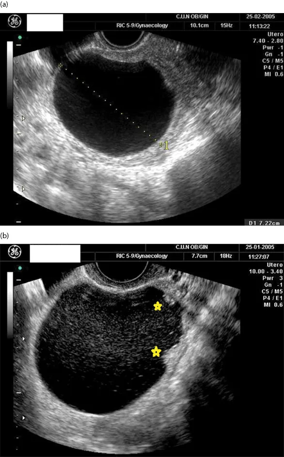
Serous cystadenoma constitutes about 25% of all benign epithelial ovarian tumors arising from the ovary; 5% of them are bilateral and most of them appear in the fourth to sixth decades of life.1 The typical ultrasound appearance of serous cystadenomas is a smooth, thin-walled, anechoic, fluid-filled lesion with a mean size of 5–8 cm (Figure 1.1a).5,6 Septations may appear in 14% of the cases and irregular wall or even papillary projections may be present in up to 3% of serous cystadenomas (Figure 1.1b).5 Color score may vary from absent to moderate flow within the cystic wall.
Mucinous Cystadenomas
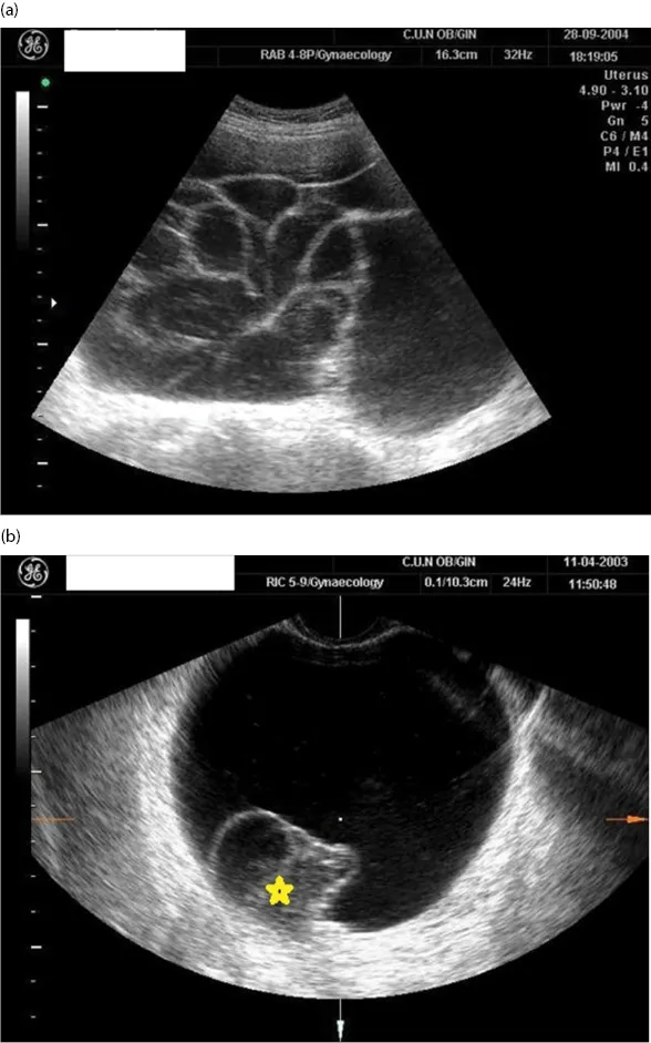
Mucinous cystadenomas constitute 25% of all benign epithelial ovarian tumors; 10% of them are bilateral and usually appear in the fifth and sixth decades of life.1 The typical ultrasound appearance of mucinous cystadenoma is a multilocular smooth cyst with a mean tumor size of 11 cm (range 3–30 cm) (Figure 1.2).7,8 Color score is usually absent, or scanty and papillary projections may appear in 20% of the cases. Caspi et al. described that the presence of different echogenicity in different locules of the lesion is almost pathognomonic of a mucinous tumor.9
Endometrioma
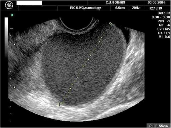
Endometrioma is addressed in detail in another chapter of the book. But know that the typical ultrasound appearance of endometrioma is a unilocular cyst with ground-glass echogenicity (Figure 1.3).5,6
Cystadenofibroma
Cystadenofibromas are relatively uncomm...
Table of contents
- Cover
- Title Page
- Copyright Page
- Contents
- Contributors
- Chapter 1: Benign Adnexal Masses and Adnexal Torsion
- Chapter 2: Pelvic Endometriosis
- Chapter 3: Ovulation, Hemorrhagic Cyst, and Hyperstimulation Syndrome
- Chapter 4: Pelvic Inflammatory Disease
- Chapter 5: Dysmenorrhea and Adenomyosis
- Chapter 6: Uterine Myoma
- Chapter 7: Müllerian Anomalies
- Chapter 8: Pelvic Congestion Syndrome and Pelvic Adhesions
- Chapter 9: Non-Gynecological Causes of Pelvic Pain
- Index
Frequently asked questions
Yes, you can cancel anytime from the Subscription tab in your account settings on the Perlego website. Your subscription will stay active until the end of your current billing period. Learn how to cancel your subscription
No, books cannot be downloaded as external files, such as PDFs, for use outside of Perlego. However, you can download books within the Perlego app for offline reading on mobile or tablet. Learn how to download books offline
Perlego offers two plans: Essential and Complete
- Essential is ideal for learners and professionals who enjoy exploring a wide range of subjects. Access the Essential Library with 800,000+ trusted titles and best-sellers across business, personal growth, and the humanities. Includes unlimited reading time and Standard Read Aloud voice.
- Complete: Perfect for advanced learners and researchers needing full, unrestricted access. Unlock 1.5M+ books across hundreds of subjects, including academic and specialized titles. The Complete Plan also includes advanced features like Premium Read Aloud and Research Assistant.
We are an online textbook subscription service, where you can get access to an entire online library for less than the price of a single book per month. With over 1.5 million books across 990+ topics, we’ve got you covered! Learn about our mission
Look out for the read-aloud symbol on your next book to see if you can listen to it. The read-aloud tool reads text aloud for you, highlighting the text as it is being read. You can pause it, speed it up and slow it down. Learn more about Read Aloud
Yes! You can use the Perlego app on both iOS and Android devices to read anytime, anywhere — even offline. Perfect for commutes or when you’re on the go.
Please note we cannot support devices running on iOS 13 and Android 7 or earlier. Learn more about using the app
Please note we cannot support devices running on iOS 13 and Android 7 or earlier. Learn more about using the app
Yes, you can access Ultrasound of Pelvic Pain in the Non-Pregnant Patient by Juan Luis Alcázar, María Ángela Pascual, Stefano Guerriero, Juan Luis Alcázar,María Ángela Pascual,Stefano Guerriero in PDF and/or ePUB format, as well as other popular books in Medicine & Family Medicine & General Practice. We have over 1.5 million books available in our catalogue for you to explore.