- 262 pages
- English
- ePUB (mobile friendly)
- Available on iOS & Android
eBook - ePub
About this book
Sonography has emerged as a substantial diagnostic tool today. This handbook aims to cover ultrasound physics, abdominal and obstetric sonography, color Doppler, high resolution sonography and USG guided interventions with multiple choice questions and case reports for practical orientation.
Trusted by 375,005 students
Access to over 1.5 million titles for a fair monthly price.
Study more efficiently using our study tools.
Information
Topic
MedicineSubtopic
Clinical MedicinePART I
USG Physics
1 Ultrasound Physics
1
Ultrasound Physics
INTRODUCTION
Ultrasound waves are defined as sound waves of high frequency that are inaudible to the ear. These are longitudinal waves that propel in a direction parallel to that of wave propagation in a medium.
High-frequency sound waves are inaudible to humans in the range of 2–20 million cycles per second (2–20 MHz)—this is the range of a diagnostic ultrasound.
Sound audible to humans is <20 KHz
Ultrasound is >20 KHz
Speed of sound in air is 330 meters per second
Speed of sound in fat is 1,450 meters per second
Speed of sound in soft tissue is 1,540–1,580 meters per second
Speed of sound in bone is 4,080 meters per second
Principle of sonography
BASED ON PULSE-ECHO PRINCIPLE
Pulses of high-frequency sound waves are transmitted to the patient. Echoes returning from various tissue boundaries are detected. The received echo produces an ultrasound image (Figure 1.1).
Electricity converted into sound—Pulse
Sound converted into electricity—Echo
If more sound is received back—suggestive of stronger reflector—whiter image
If less sound is received back—suggestive of weaker reflector—blacker image
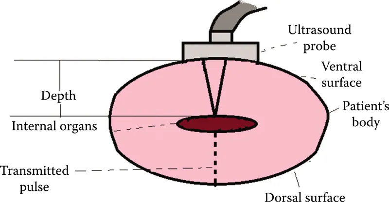
Figure 1.1 Illustrating principle of ultrasound.
Frequency: The number of cycles per second; measured in Hz (Hertz).
Wavelength: The distance between two consecutive waves. It depends on the frequency of waves and speed of propagation in the medium through which it is passing. It is inversely proportional to frequency.
Bandwidth: Range of frequencies produced by the transducer.
Pulse length: Small number of cycles in a pulse.
INSTRUMENTATION
1. Transmitter: Sends voltage to energize the transducer.
2. Transducer:
3. Receiver: To detect and amplify weak signals and send them to display It controls the dynamic range and time-gain compensation (TGC).
4. Display: To present the USG image/data in a form suitable for analysis and interpretation.
The transducer’s input is communicated to scanner through a cable and the data can be visualized on the monitor.
Following are the ways through which spatial information can be displayed:
A mode: Amplitude mode; it is used for ophthalmic purposes
B mode: Brightness mode (gray scale, real time); it is used for routine sonography
M mode: Motion mode; it is used to measure the heart rate
ULTRASONOGRAPHY TRANSDUCER
Ultrasonography (USG) transducer is a device that converts electrical energy to mechanical energy and vice versa.
It has two functions:
1. Transmitter: Electrical energy is converted to acoustic pulse, which is transmitted to the patient.
2. Receiver: Receives reflected echoes. Weak pressure changes are converted to electrical signals for processing.
It is based on the principle of piezoelectricity.
Ultrasound pulses generated by transducer are propagated, reflected, refracted, and absorbed in tissues to provide useful clinical information.
Transducers (scanning probes) are the costliest part of any ultrasound unit.
Types of transducers
The shape of the scans from different transducers is different (Figure 1.2).
1. Curved array convex transducer: Wider fan-shaped image
Useful for all body parts except echocardiography
Large versions for general abdomino-pelvic and obstetrics scan
Small high-frequency curved array scanners for transvaginal, transrectal scans
2. Linear array: Rectangular shape
Most useful for small and superficial parts such as thyroid, testicle, and breast
Vascular, musculoskeletal, and obstetric applications
3. Phased array sector scanner: Triangular fan shaped
Used in cardiac examination through intercostal scanning
Selection of transducers
The thickness of transducer (usually 0.1–1.0 millimeters) determines its frequency (inversely proportional).
Each transducer is focused at a particular depth.
Penetration of the ultrasound diminishes with an increase in frequency.
Higher the frequency, shorter the wavelength, and better the resolution.
Frequencies from 7.5 to 15 MHz are used for superficial vessels and organs such as thyroid and breast lying within—1–3 centimeters of the surface.
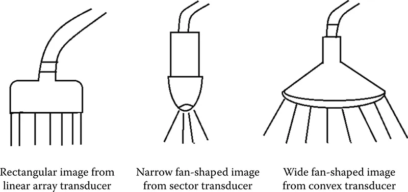
Figure 1.2 Illustrating various types of transducers.
Frequencies of 2–5 MHz are required for deeper structures in abdomen and pelvis, that is, >12–15 centimeters from the surface.
High frequency—better spatial resolution, greater attenuation, and poor penetration.
High frequencies →
• Broadens the bandwidth
• Reduces the quality factor (Q)
• Shortens the spatial pulse length (SPL)
Specialized transducers
1. Endovaginal probes for early obstetric and gynecologic problems.
2. Endorectal probes for prostate imaging.
3. Intraoperative/laparoscopic—it is used to in...
Table of contents
- Cover
- Half Title
- Title Page
- Copyright Page
- Dedication
- Table of Contents
- List of Abbreviations
- Preface
- Acknowledgments
- About the Author
- PART I USG PHYSICS
- PART II ABDOMINAL USG
- PART III OBSTETRICS USG
- PART IV COLOR DOPPLER
- PART V HIGH-RESOLUTION USG
- PART VI USG-GUIDED INTERVENTIONS
- PART VII RECENT ADVANCES IN SONOGRAPHY
- Sample Questions
- MCQs
- Answer Key
- Case Reports
- Glossary
- Index
Frequently asked questions
Yes, you can cancel anytime from the Subscription tab in your account settings on the Perlego website. Your subscription will stay active until the end of your current billing period. Learn how to cancel your subscription
No, books cannot be downloaded as external files, such as PDFs, for use outside of Perlego. However, you can download books within the Perlego app for offline reading on mobile or tablet. Learn how to download books offline
Perlego offers two plans: Essential and Complete
- Essential is ideal for learners and professionals who enjoy exploring a wide range of subjects. Access the Essential Library with 800,000+ trusted titles and best-sellers across business, personal growth, and the humanities. Includes unlimited reading time and Standard Read Aloud voice.
- Complete: Perfect for advanced learners and researchers needing full, unrestricted access. Unlock 1.5M+ books across hundreds of subjects, including academic and specialized titles. The Complete Plan also includes advanced features like Premium Read Aloud and Research Assistant.
We are an online textbook subscription service, where you can get access to an entire online library for less than the price of a single book per month. With over 1.5 million books across 990+ topics, we’ve got you covered! Learn about our mission
Look out for the read-aloud symbol on your next book to see if you can listen to it. The read-aloud tool reads text aloud for you, highlighting the text as it is being read. You can pause it, speed it up and slow it down. Learn more about Read Aloud
Yes! You can use the Perlego app on both iOS and Android devices to read anytime, anywhere — even offline. Perfect for commutes or when you’re on the go.
Please note we cannot support devices running on iOS 13 and Android 7 or earlier. Learn more about using the app
Please note we cannot support devices running on iOS 13 and Android 7 or earlier. Learn more about using the app
Yes, you can access Essentials of Abdomino-Pelvic Sonography by Swati Goyal in PDF and/or ePUB format, as well as other popular books in Medicine & Clinical Medicine. We have over 1.5 million books available in our catalogue for you to explore.