This superb, full-colour, visual revision aid has been designed specifically to candidates of the Member of the Royal College of Surgeons (MRCS) examination. It is also an invaluable diagnostic aid for all medical students and trainees, especially those on surgical rotations. Questions are based on a single clinical picture with comprehensive answers overleaf. Topics covered by this volume include laparoscopic surgery, upper and lower GI endoscopy, colorectal surgery and urology.
- 224 pages
- English
- ePUB (mobile friendly)
- Available on iOS & Android
eBook - ePub
About this book
Trusted by 375,005 students
Access to over 1.5 million titles for a fair monthly price.
Study more efficiently using our study tools.
Information
Section 1
Laparoscopic surgery
Case 1
The following pictures demonstrate the initial approach to a laparoscopic operation.
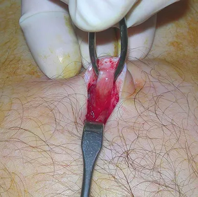
Figure 1.1a
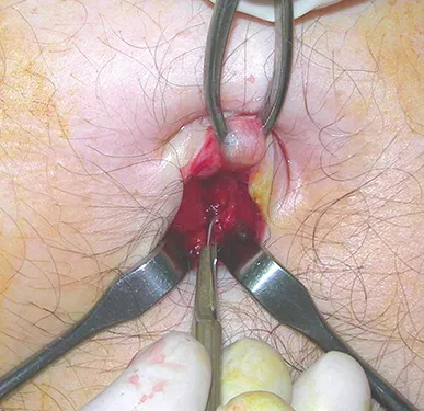
Figure 1.1b
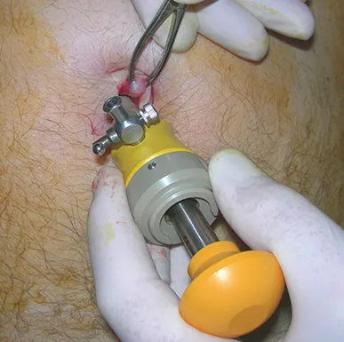
Figure 1.1c
Questions
Q1 Which technique for access to create a pneumoperitoneum is shown in Figures 1.1a–c?
Q2 Which anatomical structures need to be identified when applying it?
Q3 What alternative technique was widely used before this access?
Answers
A1 The Hasson, or ‘modified’ Hasson technique.
A2 The umbilicus and its insertion into the linea alba. The incision is made at this point, and the peritoneum punctured under direct vision to gain intraperitoneal access. A blunt trocar is then inserted and the abdomen insufflated with CO2 under direct vision.
A3 The Veress needle puncture. This is still used in some centres but concern has been raised over its technique of ‘blind’ puncture, which can result in increased risk of bowel or vascular trauma.
Case 2
Figure 1.2 shows part of a stack used in laparoscopic surgery, the ‘endoflator’. It is important to know how this equipment works.
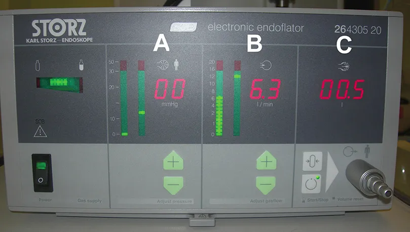
Figure 1.2
Questions
Q1 Describe the function of each of the columns labelled A, B, C.
Q2 What does the display to the left of ‘A’ indicate?
Q3 What might cause the left-hand LED in ‘A’ to rise sharply during surgery?
Answers
A1 This unit controls CO2 insufflation. ‘A’ indicates the pressure level within the abdomen and allows the constant pressure to be set at a particular limit. Here it is set at 12 mmHg (right-hand LED). The actual abdominal pressure is indicated by the left-hand LED, zero at present as the unit is disconnected. ‘B’ indicates gas flow rate, usually set at 6–10 Litres/minute. Once the abdomen has reached its preset pressure the flow rate falls to zero. When gas escapes, reducing the pressure, this is detected by the unit and the flow restarts. ‘C’ indicates the total amount of gas used.
A2 This shows how much gas remains in the cylinder, and turns red when it is running low.
A3 A sudden rise would be accompanied by an alarm signal, and indicates a sudden rise in the intra-abdominal pressure. A common cause of this is the patient beginning to wake up with the return of voluntary abdominal wall contraction (this is often an earlier ‘warning signal’ for the anaesthetist than the anaesthetic machine responses!). It may also happen with a mechanical block to the gas pipe (e.g. the tap is turned off).
Case 3
A diagnostic laparoscopy was undertaken for acute-onset right upper abdominal pain in a 17-year-old female patient. The ultrasound scan was normal.
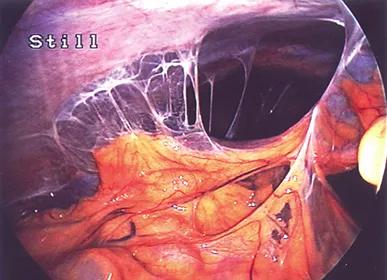
Figure 1.3
Questions
Q1 Figure 1.3 shows the view towards the right lobe of the liver. What pathology is shown on the picture?
Q2 What underlying disease is likely to have caused it?
Q3 What risks are associated with this condition?
Q4 What is the appropriate management for this...
Table of contents
- Cover
- Half Title
- Title Page
- Copyright Page
- Contents
- Foreword
- Preface
- List of contributors
- Acknowledgements
- Dedications
- Section 1: Laparoscopic surgery
- Section 2: Gl endoscopy
- Section 3: Colorectal surgery
- Section 4: Urology
- Section 5: Radiology for the surgical trainee
- Reference
- Index
Frequently asked questions
Yes, you can cancel anytime from the Subscription tab in your account settings on the Perlego website. Your subscription will stay active until the end of your current billing period. Learn how to cancel your subscription
No, books cannot be downloaded as external files, such as PDFs, for use outside of Perlego. However, you can download books within the Perlego app for offline reading on mobile or tablet. Learn how to download books offline
Perlego offers two plans: Essential and Complete
- Essential is ideal for learners and professionals who enjoy exploring a wide range of subjects. Access the Essential Library with 800,000+ trusted titles and best-sellers across business, personal growth, and the humanities. Includes unlimited reading time and Standard Read Aloud voice.
- Complete: Perfect for advanced learners and researchers needing full, unrestricted access. Unlock 1.5M+ books across hundreds of subjects, including academic and specialized titles. The Complete Plan also includes advanced features like Premium Read Aloud and Research Assistant.
We are an online textbook subscription service, where you can get access to an entire online library for less than the price of a single book per month. With over 1.5 million books across 990+ topics, we’ve got you covered! Learn about our mission
Look out for the read-aloud symbol on your next book to see if you can listen to it. The read-aloud tool reads text aloud for you, highlighting the text as it is being read. You can pause it, speed it up and slow it down. Learn more about Read Aloud
Yes! You can use the Perlego app on both iOS and Android devices to read anytime, anywhere — even offline. Perfect for commutes or when you’re on the go.
Please note we cannot support devices running on iOS 13 and Android 7 or earlier. Learn more about using the app
Please note we cannot support devices running on iOS 13 and Android 7 or earlier. Learn more about using the app
Yes, you can access MRCS Picture Questions by Tjun Tang,Bandipalyam Vamana Rao Praveen in PDF and/or ePUB format, as well as other popular books in Medicine & Medical Theory, Practice & Reference. We have over 1.5 million books available in our catalogue for you to explore.