![]()
PART 1
1 | History |
2 | Mesenteric and peritoneal anatomy |
3 | Embryologic development of the mesentery, peritoneal reflection, and Toldt’s fascia |
4 | Histology of the mesentery |
5 | Toldt’s fascia |
6 | Mesenteric physiology |
7 | Pathology of the mesentery |
8 | Radiographic appearance of the mesentery and peritoneum |
9 | Operative nomenclature |
10 | Teaching mesenteric principles |
11 | Gastroenterology |
![]()
1
History
J. CALVIN COFFEY AND NICOLA O’RIORDAN
Introduction | |
Carl Toldt | |
Sir Frederick Treves | |
Limited support for Toldt’s observations | |
Radiology | |
Renaissance in focus on the mesentery | |
Laparoscopic and robotic surgery: The craft of colorectal surgery | |
Anatomic continuity: A simpler principle | |
Future directions | |
Summary | |
References | |
Two roads diverged in a wood, and I—
I took the one less travelled by,
And that has made all the difference.
Robert Frost
INTRODUCTION
For centuries, the mesentery and associated peritoneal lining have been regarded as structurally complex. In 1885, Sir Frederick Treves provided the first comprehensive description of both, emphasizing that while some mesenteric regions persisted in adulthood, others regressed and were lost [1]. For example, the small intestinal mesentery, transverse, and sigmoid mesocolon were consistently identifiable in adults, while the right and left mesocolon were identifiable in a minority only. Treves’ descriptions were welcome at the time, given the apparent complexity of the topic, and were subsequently indoctrinated in virtually all anatomic, embryologic, clinical, and related literature [1–3]. To the present, the first chapter of most reference texts on intestinal surgery focuses on anatomy and physiology and is based on Treves’ descriptions. A review of later chapters dealing with techniques in intestinal removal identifies a remarkable disparity. The right and left mesocolon are invariably present in the adult and must be resected like any other mesenteric region. Put simply, intestinal surgery has always relied on the persistence of all regions of the mesentery.
Numerous factors contributed to the divergence of anatomic and surgical approaches to the mesentery and peritoneum. Since the time of Treves’ anatomic-based research, surgeons focused increasingly on cellular aspects of disease. With increasing awareness of the molecular basis of surgical disease, the emphasis of research shifted away from the anatomic-based craft component. More recently, laparoscopic and robotic surgery have increased focus on the “craft” component of surgery. In keeping with this, the field of surgical anatomy has increased in relative significance and led to the demonstration of continuity of the mesenteric organ from the small intestinal mesentery to the mesorectum [4,5]. The following chapter will demonstrate these shifting trends and clarify the manner in which recent demonstrations allow a reconciliation of anatomic and surgical approaches to this important organ. This chapter finishes by demonstrating the opportunities that now occur across a broad array of clinical and non-clinical sciences.
CARL TOLDT (1840–1920)
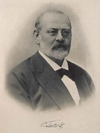
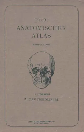
Carl Florian Toldt was born on May 3, 1840, in Bruneck, Austria. After spending much of his childhood repairing clocks, he received his doctorate in 1864 at St. Joseph’s University in Vienna and was appointed Professor of Anatomy at the University of Vienna in 1875 (Figure 1.1). He became Professor of Anatomy at the German university in Prague. He subsequently returned to Vienna in 1884 to work with his colleague, Langer, and together they established the Anatomy Institute of Vienna. Carl Toldt’s best-known anatomic work was Anatomischer Atlas für Studierende und Aerzte (An Atlas of Human Anatomy for Students and Physicians) (Figure 1.2), which was translated into English. Despite the superb quality of this work, and its anatomic accuracy, it has been little referenced overall. Toldt died from pneumonia in Vienna in November 1920 [6–9].
Toldt’s descriptions were based on dissection of fresh cadavers that had not been exposed to corrosive preservative agents. He first observed that intestinal mesenteries did not simply comprise two thin layers of closely apposed cells (i.e., the mesothelia) but rather contained vessels, nerves, and fat. He called the resultant complex of structures the “lamina mesenteria propria” (Figure 1.3). While Treves described disappearance of the right and left mesocolon as humans matured into adults, Toldt maintained they persisted and attached (i.e., flattened against) to the abdominal wall. Where they attached, Toldt identified a thin seam of connective tissue (Toldt’s fascia) separating mesentery from abdominal wall, just as two layers of rock might be separated by a seam of rock (see Chapter 2). Toldt suggested that wherever the mesentery attached to the abdominal wall, the cellular layer lining both (i.e., the mesothelium) underwent a “gradual disappearance” to “admit contact and fusion of their connective tissue laminae.” Toldt also suggested that the outer cellular layer of the intestine, the tunica serosa, could undergo a similar process and merge with the covering mesothelium of adjacent mesentery. There is a striking similarity between current descriptions, and those of Toldt, and it is remarkable that his work should have been so infrequently referenced over the past two centuries [6–9].
Figure 1.1 Carl Toldt (1840–1920).
Figure 1.2 Cover illustration of Anatomischer Atlas Fur Studierende und Aertze.
SIR FREDERICK TREVES
Frederick Treves was born in Dorset, England, in 1853 (Figure 1.4). He received his medical education at the London School of Medicine and became assistant surgeon at the London hospital in 1879. In 1883 he was appointed as surgeon and head of the department of Anatomy. He famously housed Joseph Merrick, “the Elephant Man,” in his attic until Merrick died in 1890 [6,10]. Treves was awarded the Jacksonian prize for dissertations on the pathology, diagnosis, and treatment of obstruction of the intestine and numerous Hunterian lectures on the anatomy of the intestinal canal and the peritoneum. He served in the Boer war in 1899. He was knighted by King Edward VII on whom he performed an appendectomy in 1902. He was a noted travel writer and took up final residence in Geneva (Switzerland) due to poor health. He died of peritonitis in 1923 [6,10,11].
Treves described the human mesentery as fragmented. Accordingly, the right and left mesocolic components of the mesentery are, according to Treves, mostly absent in the adult human. He described the small intestinal, transverse, and sigmoid mesentery as persisting into adulthood and attaching directly to the abdominal wall (Figure 1.5). At the time, his descriptions provided a welcome rationalization of what was, and still is, regarded as a complex anatomic topic (i.e., mesenteric and peritoneal anatomy in the adult human). Although some aspects of his descriptions of the mesentery and peritoneum are now regarded as inaccurate, he was correct in describing a “mesenteric root” at the origin of the superior mesenteric artery. He was also correct in describing the mesentery of the appendix as arising from the undersurface of the mesentery in the right iliac fossa. Treves’ stunning descriptions were made at a time when significant advances were occurring in anatomic and safe surgery, a factor that is likely to have aided in their indoctrination in mainstream literature....