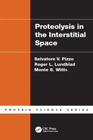
Proteolysis in the Interstitial Space
- 297 pages
- English
- ePUB (mobile friendly)
- Available on iOS & Android
Proteolysis in the Interstitial Space
About this book
Most clinical laboratory tests utilize interstitial and extravascular such as blood, urine, cerebral spinal fluid (CSF), and saliva. For example, CSF is monitored in the context of cancer for both diagnostic and therapeutic reasons. And yet, our understanding of the makeup of interstitial fluids, their relationships to disease, as well as their commercial importance in therapeutics and diagnostics remains rudimentary. Although sometimes perceived as static, interstitial and extravascular fluids are surprisingly dynamic. More than half of serum albumin is in the extravascular space. These fluids move rapidly between the intravascular and extravascular spaces - one entire plasma volume is exchanged very nine hours. In the first half of the book, the authors cover fundamental concepts of interstitial fluids, including their composition and function. They then further review the mechanisms by which interstitial fluids are regulated, characterizing the importance of hyaluronan – a major constituent of interstitial spaces and an a component of synovial fluid; and, outlining the regulation of proteolysis in the interstitial space. In the second half of the book, the authors focus on the coagulation system. This system has been studied extensively in the context of vascular spaces. But many of its components exist in the interstitial spaces. Chapters are devoted to the fibrinolytic system, kallikrein, matrix metalloproteinases, coagulation factors, and protease inhibitors – all are interstitial. By covering a unique array of topics with broad application to biomedical scientists, this book expands our understanding of the importance of interstitial spaces and the fluids that move through and reside in this extravascular environment.
Tools to learn more effectively

Saving Books

Keyword Search

Annotating Text

Listen to it instead
Information
1 | Composition and Function of the Interstitial Fluid |
Protein Content of Various Human Body Fluids and Secretions
Fluid | Protein (mg/mL)a | Comment | Refs. |
Extracellular fluid | N/A | The body fluid can be divided into two major components: the intracellular fluid and the extracellular fluid. Between 60% and 70% of the body fluid is intracellular in nature, while the remainder is extracellular in nature. The extracellular fluid, in turn, consists of two primary components: intravascular fluid (blood plasma; approximately 25%) and extravascular fluid (approximately 75%). The extravascular fluid consists mostly of interstitial fluid with small specialized fluids in various spaces; specialized fluids include cerebrospinal fluid, synovial fluid, and ocular fluid. | 1–5 |
Blood plasma | 78.9 ± 0.5b | A protein-rich fluid defined by being confined within the vascular system and representing one-quarter to one-fifth of the total extracellular fluid. It is in equilibrium with the interstitial fluid, which feeds into the lymphatic system and is returned to the venous system. Other areas of extracellular fluid include the peritoneal fluid, ocular fluid, and cerebrospinal fluid. Plasma is also defined as the protein-rich fluid obtained by the removal of the cellular elements of whole blood collected with an anticoagulant. | 6–8 |
Blood serum | 72.9 ± 0.5b | A protein-rich fluid derived from the clotting of plasma or whole blood. Most frequently collected by the clotting of whole blood collected without the addition of an anticoagulant. The protein concentration of serum is usually less that of corresponding plasma, reflecting the loss of fibrinogen and other plasma proteins. Serum may also contain products secreted by platelets and other cellular elements during the process of coagulation. | 6–8 |
Interstitial fluidc | 50.9d | The concentration of protein in interstitial fluid is 40–60% of that in plasma. The volume of interstitial fluid is two to three times larger than plasma volume and the concentration of a given protein in the interstitial fluid depends on the excluded interstitial volume for a specific protein and the size of the protein. Albumin is the most common protein in interstitial fluid, with lower concentrations of larger proteins such as IgG. | 9–11 |
Interstitial fluid | 29.8 | Plasma protein concentration was given at 70.0 mg/mL; the interstitial volume was 8.4 L, with an excluded interstitial volume of 2.1 L. | 12 |
Interstitial fluid | 27.2 18.3 | Wick fluid.e Blister technique.e | 13 |
| Interstitial fluid | 37f 24 | Perivascular. Peribronchial. | 14 |
Lymph | 26–51e | Lymph is derived from plasma via interstitial fluid. There is tissue variability in lymph flow rate and regional composition. In this study, as with others, albumin was the major protein. This study also referred to other proteins as members of the classical globulin fractions. | 15 |
Lymph | 17–25f | Protein concentration increased to 44 mg/mL in skin lymph but not muscle lymph after thermal injury. | 16 |
Lymphg | 42 | The lymph/plasma ratio was 0.71 for total protein, 0.70 for albumin, and 0.25 for immunoglobulin. | 17 |
Lymphh | 25–27 | The protein concentration of lymph was slightly less than that of interstitial fluid. The concentration of lactic dehydrogenase was much higher in interstitial fluid than in lymph; the concentration in lymph in turn is much higher than that in plasma. | 18 |
Lymphi | 27 | The protein concentration of lymph was slightly less than that obtained for interstitial fluid (25 mg/mL). Albumin is the major protein (18 mg/mL), with smaller quantities of globulinj (8.2 mg/mL). | 19 |
Peritoneal fluid | 25 | Peritoneal fluid is usually obtained only the in the case of ascites; it is labeled a transudate if the protein concentration is less than 25 mg/mL and an exudate if the protein concentration is greater than 25 mg/mL.k A transudate can result from increased hydrostatic pressure, while an exudate can result from decreased capillary permeability. | 20–22 |
Peritoneal fluid | 42.2 ± 6 | Peritoneal fluid obtained from norma... |
Table of contents
- Cover
- Half Title
- Title Page
- Copyright Page
- Dedication
- Table of Contents
- Preface
- Acknowledgments
- Authors
- Chapter 1 Composition and Function of the Interstitial Fluid
- Chapter 2 The Extracellular Matrix, Basement Membrane, and Glycocalyx
- Chapter 3 The Biochemistry of Hyaluronan in the Interstitial Space
- Chapter 4 Proteolysis in the Interstitium
- Chapter 5 The Fibrinolytic System in the Interstitial Space
- Chapter 6 Kallikrein in the Interstitial Space
- Chapter 7 Matrix Metalloproteinases in the Interstitial Space
- Chapter 8 Coagulation Factors in the Interstitial Space
- Chapter 9 Protease Inhibitors in the Interstitial Space
- Index
Frequently asked questions
- Essential is ideal for learners and professionals who enjoy exploring a wide range of subjects. Access the Essential Library with 800,000+ trusted titles and best-sellers across business, personal growth, and the humanities. Includes unlimited reading time and Standard Read Aloud voice.
- Complete: Perfect for advanced learners and researchers needing full, unrestricted access. Unlock 1.4M+ books across hundreds of subjects, including academic and specialized titles. The Complete Plan also includes advanced features like Premium Read Aloud and Research Assistant.
Please note we cannot support devices running on iOS 13 and Android 7 or earlier. Learn more about using the app