![]()
1 Friction Ridge Skin and Prints
The aim of this chapter is to provide a summary of the basic elements of friction ridge skin morphogenesis and their relationship to friction ridge skin variability. More extensive accounts for fingermark examiners can be found in the literature (Ashbaugh 1999; Bush 2002; Wertheim and Maceo 2002; Maceo 2011; Wertheim 2011). These contributions, with their associated references, constitute the essential material required to gain an understanding of the biological basis for friction ridge pattern variability. They complement and extend the work undertaken by earlier pioneers such as Wilder and Wentworth (1932) and Cummins and Midlo (1961).
Two cornerstones to the use of fingerprints as a means of personal identification are the overall permanence (a.k.a. persistency, durability, or reproducibility) and the high selectivity (a.k.a. discrimination) of friction ridge skin. It is thanks to these two attributes, and to the fact that fingerprints can be classified with relative ease, that the technique imposed itself over precedent identification methods based on anthropometric measurements. Both of these foundations— permanence and selectivity—have been challenged and confirmed through 100 years of fingerprint identification practice and their scientific foundations lie within biological research.
Unless examiners have a good understanding of friction ridge skin morphogenesis (the biological development of form), the basic tenet for individuality is, unfortunately, often resolved by using standard, shallow statements such as “nature never repeats itself” (McRoberts 1996). We strive to avoid a justification for individualization that only revolves around the tautological argument that every entity in nature is unique. The permanence and selectivity of friction ridge skin should be fully understood from a biological perspective and then applied in assessing fingermark comparisons.
1.1 STRUCTURE OF THE SKIN
Skin is an essential organ of the human body. Finger, palm, and sole areas of the epidermis display a series of friction ridges taking various forms and shapes. These volar areas of the skin are known to display friction ridge skin. Depending on the surface considered, we generally refer to them as fingerprints, palm prints, and soleprints. It is postulated that the essential function of having friction ridge skin is to increase grip.
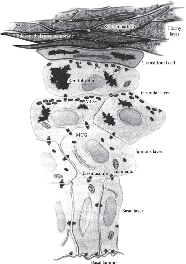
The skin is usually divided into two distinct layers. The outer layer (Figure 1.1), called the epidermis, is a stratified epithelium of five sublayers, listed as follows from bottom to top:
Basal generating layer (stratum germinativum)
Spinous layer (stratum spinosum)
Granular layer (stratum granulosum)
Transitional hyalin layer (stratum lucidum)
Horny cornified layer (stratum corneum)
The layers of the epidermis are named on the basis of microscopic shape of the keratinocyte cells that constitute them. The layer under the epidermis is called the dermis and is 15–40 times thicker than the epidermis and constitutes the primary mass of the skin. The cornified layer exposed to the environment is made up of 15–20 layers of flat dead cells that are regularly shed through abrasion and replaced by keratinization. All these cells originate from initial cuboidal-shaped cells formed on the basal layer (cells just above the basal lamina) that migrate through the epidermal layers up to the horny layer. The cells move upward simultaneously with surrounding cells. The basal cells do not migrate and remain firmly attached to the generating layer. During this process, cells change shape, reduce their activity, and take up keratin (a water-repellent protein). All cells of the epidermis therefore originate from the basal layer at the interstice between the dermis and the epidermis. As we will explain in more details later, permanency of the friction ridge pattern is largely due to this generative process, whereby the cells constituting the epidermis (and thus its shapes) are produced on the inner protected basal layer just above the dermis. Only damage to the basal layer will result in permanent scars on the epidermis.
FIGURE 1.1 Structure of the epidermis from basal lamina to horny layer. (Reproduced from Montagna, W. and Parakkal, P.F., The Structure and Function of Skin, 3rd ed., Academic Press, London, U.K., 1974. With permission.)
1.2 MORPHOGENESIS OF FRICTION RIDGE SKIN: PRIMARY DERMAL RIDGE DEVELOPMENT
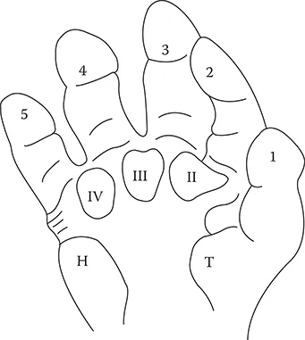
The morphogenesis of friction ridge skin starts during the very first weeks of gestation. In fact, we can only speak of estimated gestational age (EGA). The hand starts to develop from 5 to 6 weeks EGA. The first fingers appear around 6–7 weeks. At that time, volar pads appear on the palm (interdigital pads first, followed by thenar and hypothenar pads). Volar pads are transient swellings of mesenchymal tissue under the epidermis on the volar surfaces of the fetus. Volar pads appear on each finger at 7–8 weeks (Figure 1.2). These pads remain visible until 10 weeks, when the growth of the hand overtakes the pads, rendering them not visible by week 16 EGA. This phenomenon is often described as the “regression” stage of the volar pads. It is between weeks 11 and 20 that the major development of friction ridge skin occurs. The volar pads provide the bedding for that development.
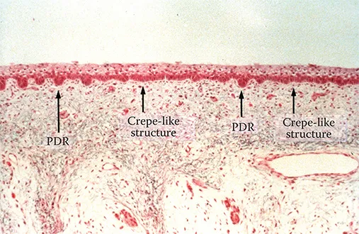
At around 10 weeks EGA, cells on the basal layer start to proliferate. Prior to ridge development, the embryonic epidermal surface—the periderm—is three or four cell layers thick and smooth on its outer surface (Figure 1.3). The location of the initial proliferations seems coincident with sweat gland development, but it could also be associated with the arrangement of superficial dermal nerves (Merkel cell clusters and Meissner corpuscles) organized in an approximately two- dimensional hexagonal grid that orchestrates the spacing and general arrangement of the papillary ridges (Dell and Munger 1986). These cells, each associated with a sweat gland, multiply rapidly and fuse into ridges called “ledges” (Hale 1952). These ridges, called primary dermal ridges (PDRs), are still immature and will start to mature by developing downward within the dermis. Individual dermal ridges are not yet differentiated on the areas surrounding these focal areas; rather, the dermis presents a primordial crepe-like appearance (Figure 1.3). The latter is predictive of the basic orientation of the ridge structure to be manifested there later (Okajima and Newell-Morris 1988).
The first obvious manifestation of friction ridge skin is primary ridges on the dermis with fully formed minutiæ (ridge endings or bifurcations). The configuration can be viewed as a series of ridge units (each associated with a sweat pore) that have fused together into ridges of various lengths— lengths being defined by the number of ridge units between two minutiæ, the smallest ridge being a single ridge unit.
FIGURE 1.2 Volar pads as they appear on the surface of the hand. The first to appear are the second, third, and fourth interdigital pads (II to IV), followed by the apex finger pads. (Reproduced from Ashbaugh, D.R., in Qualitative-Quantitative Friction Ridge Analysis—An Introduction to Basic and Advanced Ridgeology, CRC Press, Boca Raton, FL, 1999. Figure 2.20. With permission.)
FIGURE 1.3 Cross-sectional view of the skin of a fetus at the initial stages of primary ridge development, showing the primary dermal ridge and the crepe-like structure. (Image obtained from M. Okajima, Tokyo, Japan. With permission.)
When the surface of the dermis is examined at its various stages of development, ridges start to be visible from the apex of the volar pad (core of the fingerprint), the distal periphery (tip of the finger), and the distal interphalangeal flexion crease area. These three fronts of ridges develop at different speeds until they ultimately converge to fully cover the glabrous dermal surface (Figures 1.4 through 1.6). Such pattern development may follow the molecular mechanism proposed by (Turing 1952), called the reaction–diffusion system, which can develop periodic patterns from an initially homogeneous state. Many models—as will be discussed later—have since been proposed in mathematical biology to account for patterning phenomena in morphogenesis based on this mechanism, and a computer model has been used to generate fingerprint patterns.
FIGURE 1.4 Image of the dermal surface of the index finger of a fetus (11 weeks estimated gestational age). Note the initial development of the ridges on the apex of the finger. Dark staining indicates primary ridges. (Image obtained from M. Okajima, Tokyo, Japan. With permission.)
FIGURE 1.5 Dermal surface of the index finger of a fetus (11 weeks estimated gestational age), showing the development of ridges from various development fronts. Dark staining indicates primary ridges. (Image obtained from M. Okajima, Tokyo, Japan. With permission.)
FIGURE 1.6 Dermal surface of the index finger of a fetus (14 weeks estimated gestational age), showing the final friction ridge pattern. (Image obtained from M. Okajima, Tokyo, Japan. With permission.)
1.3 FACTORS AFFECTING THE GENERAL PATTERN AND THE CONFIGURATION OF MINUTIÆ
The general pattern taken by the ridges is dependent on the following interrelated factors (Bonnevie 1924; Cummins 1926; Penrose and Loesch 1969; Babler 1987):
Shape (symmetry) and size of the volar pads (Penrose hypothesized that the flow of the ridges follows the lines of curvature of the skin on the volar pad) (Penrose and O’Hara 1973); research in mathematical biology confirmed this hypothesis (Smith 1979; Mardia et al. 1992)
Timing between the regression of the volar pads and the onset of primary ridge formation
Relative speed of the development fronts
Bone morphology (Babler 1991)
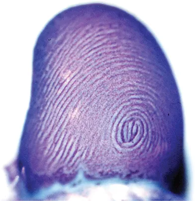
It is important to recognize that the basic form of the general pattern is established before the initial development of the PDR (Figure 1.7). Wertheim (2011), based on Wertheim and Maceo (2002), provided detailed examples of the various general patterns associated with the behavior of the aforementioned variables, with didactic animations at the following website (accessed January 31, 2016): http://www.clpex.com/animation.htm.
Symmetrical pads will tend to develop into whorls; displaced laterally volar pads will lead to result in loops (to the right or to the left depending on the leaning of the pad). Very weakly elevated pads will produce arches. The pattern size that can be measured by the distance between the core of the pattern and the delta(s) depends on the overall size of the pad at the moment where friction ridges started to develop. The timing of the onset of volar pad regression and the timing of the onset of primary ridge formation are critical factors affecting the overall size of the fingerprint pattern. Hence, timing and symmetry control of the volar pads, both conditioned on the aforementioned interrelated factors, are driving the overall ridge flow.
Recent work by Kücken (2004, 2007) and Kücken and Newell (2004, 2005) in relation to the formation of the general patterns of friction ridge skin brings a very comprehensive bibliographical resource and new insights on the morphogenesi...