- 339 pages
- English
- ePUB (mobile friendly)
- Available on iOS & Android
About This Book
This fully illustrated text is an essential guide for trainees in Haematology and Medicine studying for higher examinations, as well as for professionals wishing to expand their knowledge or revalidate.
The book contains over 100 illustrated cases covering the whole field of malignant and non-malignant haematology, including coagulation problems and infectious diseases. Each case contains a set of questions written by two Royal College examiners, with answers on the reverse page. Readers can make differential diagnoses and devise treatment plans and prognoses, before turning the page to read the experts' detailed answers.
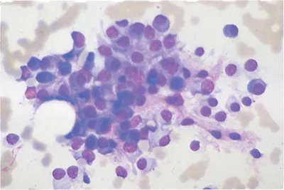
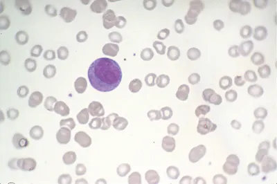
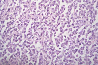
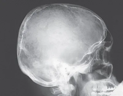
The cases are presented in random order – just as they would be in real life – and are of varying lengths and degrees of difficulty, accompanied by hundreds of colour photomicrographs, photographs, and x-rays.
This new edition is revised and updated, with new cases, images, and tables.
Frequently asked questions
Information
Section 1
GENERAL AND MALIGNANT HAEMATOLOGY
CASE 1
QUESTIONS
Haemoglobin (Hb) | 71 g/L |
White blood cells (WBC) | 4.6 × 10⁹/L |
Platelets | 112 × 109/L |
Urea | 46 mmol/L |
Creatinine | 905 mmol/L |
Ca2+ | 3.60 mmol/L (N 2.1–2.6 mmol/L) |
Albumin | 26 g/L (N 35–42 g/L) |
Total protein | 120 g/L (N 65–80 g/L) |
Alkaline phosphatase | 143 U/L (N 30–130 U/L) |
Uric acid | 0.48 mmol/L (N 0.3–0.4 mmol/L) |
Q1.i | Comment on the above results. |
Q1.ii | Comment on the bone marrow aspirate. |
Case 1: ANSWERS
A1.i | The results indicate anaemia and thrombocytopenia with marked renal failure. Hypercalcaemia with normal alkaline phosphatase suggests primary bone marrow malignancy. The raised total protein suggests myeloma. |
A1.ii | The bone marrow is infiltrated by plasma cells, confirming myeloma. Plasma cell leukaemia is an aggressive form of myeloma characterised by large numbers of circulating plasma cells. |
Q1.iii | Comment on the aspirate of this patient’s skin nodule. |
Q1.iv | Comment on the skull x-ray. |
Q1.v | What is the diagnosis? |
Q1.vi | How should he be treated? |
A1.iii | The skin deposit is also due to myeloma infiltration. |
A1.iv | The skull x-ray shows multiple lytic lesions, a characteristic finding in myeloma. |
A1.v | Myeloma. |
A1.vi | The hypercalcaemia and renal failure require urgent therapy with rehydration to promote diuresis. An intravenous urogram (IVU) should not be done as the patient should not be dehydrated. However, abdominal ultrasound scan to exclude renal obstruction is valuable in acute renal failure. |
Baseline tests should include paraprotein quantification in serum and urine (Bence Jones protein), skeletal survey, beta-2 microglobulin and C-reactive protein. Other baseline tests should include a coagulation profile, culture of mid-stream urine and assessment of antibodies to hepatitis A, B and C. Serumfree light chains should be estimated as they are elaborated by the tumour cells and are often the principal cause of the renal toxicity. A renal biopsy should be considered, and the nephrologist will wish to undertake a range of tests to exclude other causes of acute renal impairment. A complete cardiac assessment including echocardiography is important, and the presence of amyloid deposition, for example, in heart and kidneys should be considered. ... |
Table of contents
- Cover
- Half Title
- Title Page
- Copyright Page
- Contents
- Foreword
- Acknowledgements
- List of Abbreviations
- Section I: General and malignant haematology
- Section II: Coagulation
- Section III: Quality control
- Index