Hypopigmentation
  1. 180 pages
  2. English
  3. ePUB (mobile friendly)
  4. Available on iOS & Android
eBook - ePub
Book details
Book preview
Table of contents
Citations

About This Book

There are many disorders of a lack of pigmentation in the skin, with different causations

and effects, of which vitiligo is only the best known; this comprehensive text from international

experts will enable clinicians to diagnose the full range of these conditions and suggest

the most effective management options for their patients.

Contents: Basic concepts of melanocyte biology * Approach to hypopigmentation *

Historical review of vitiligo * Epidemiology and classification of vitiligo * Pathophysiology

of vitiligo * Segmental vitiligo * Childhood versus post-childhood vitiligo * Pharmacological

therapy of vitiligo * Surgical treatment of vitiligo * Phototherapy and lasers in the treatment

of vitiligo * Emerging treatments for vitiligo * Tuberous sclerosis complex * Oculocutaneous

albinism * Hermansky-Pudlak syndrome, Chediak-Chigasi syndrome, and Griscelli

syndrome * Piebaldism * Waardenburg syndrome * Alezzandrini syndrome, Margolis

syndrome, Cross syndrome, and other rare genetic disorders * Mosaic hypopigmentation

* Skin disorders causing post-inflammatory hypopigmentation * Infectious and parasitic

causes of hypopigmentation * Melanoma leukoderma * Halo nevi * Drug-induced hypopigmentation

* Hypopigmentation from chemical and physical agents * Guttate hypomelanosis

and progressive hypomelanosis of the trunk (progressive macular hypomelanosis)

Frequently asked questions

Simply head over to the account section in settings and click on ā€œCancel Subscriptionā€ - itā€™s as simple as that. After you cancel, your membership will stay active for the remainder of the time youā€™ve paid for. Learn more here.
At the moment all of our mobile-responsive ePub books are available to download via the app. Most of our PDFs are also available to download and we're working on making the final remaining ones downloadable now. Learn more here.
Both plans give you full access to the library and all of Perlegoā€™s features. The only differences are the price and subscription period: With the annual plan youā€™ll save around 30% compared to 12 months on the monthly plan.
We are an online textbook subscription service, where you can get access to an entire online library for less than the price of a single book per month. With over 1 million books across 1000+ topics, weā€™ve got you covered! Learn more here.
Look out for the read-aloud symbol on your next book to see if you can listen to it. The read-aloud tool reads text aloud for you, highlighting the text as it is being read. You can pause it, speed it up and slow it down. Learn more here.
Yes, you can access Hypopigmentation by Electra Nicolaidou, Clio Dessinioti, Andreas Katsambas, Electra Nicolaidou, Clio Dessinioti, Andreas Katsambas in PDF and/or ePUB format, as well as other popular books in Medicine & Medical Theory, Practice & Reference. We have over one million books available in our catalogue for you to explore.

Information

Publisher
CRC Press
Year
2019
ISBN
9781351379625
1
Basic concepts on melanocyte biology
Mauro Picardo and Daniela Kovacs
Contents
Evolution/adaptation of human pigmentation and its heterogeneity
Melanocytes and melanin synthesis
Melanosome transport inside melanocytes and melanosome transfer to keratinocytes
Cellā€“cell crosstalk in the control of melanocyte functionality
Extracellular matrix microenvironment and melanocyte homeostasis
Concluding remarks
References
Evolution/adaptation of human pigmentation and its heterogeneity
Melanocytes participate as the major performers in the complex scenario of biological components regulating the process of pigmentation. These cells are specialized in the synthesis of melanin pigments inside membrane-bound organelles, the melanosomes. Along with hemoglobin and carotenoids, melanin is the main pigment responsible for the color variations of our skin and hair. Differences in skin and hair color are considered to be adaptive responses and to be highly related to ultraviolet (UV) exposure and latitude. During evolution, human ancestors living in equatorial Africa were probably characterized by light pigmentation of the body, which was, however, covered by dark hair. The gradual loss of body hair paralleled the increase in the epidermal and stratum corneum thickness and in dark-photoprotective eumelanin pigmentation to prevent the damages of ultraviolet radiation (UVR) near the equator. Under intense UVR exposure, dark skin developed as a protective mechanism to limit destruction of cutaneous and systemic folate. Folate regulates important biological processes such as DNA synthesis, repair, methylation, and maintenance of active spermatogenesis, as well as melanin production. Folate deficiency has been linked to pregnancy complications and severe fetal abnormalities in neural tube development. The sensitivity of folate and of its main serum form, 5-methyltetrahydrofolate, to be degraded by UVR and reactive oxygen species (ROS) supports the hypothesis according to which the increased pigmentation occurring in high UVR-exposed terrestrial areas evolved to prevent fertility reduction caused by folate photodegradation. As hominins gradually moved outside of tropical latitudes, toward Eurasia, the Americas, and nonequatorial Africa, the intensity and duration patterns of UV exposure decreased together with a reduced potential for vitamin D production, thus favoring the promotion of depigmentation. Therefore, the wide array of pigmentation characterizing modern humans seems to be guided on the one hand by the need to promote photoprotection near the equator (stimulating the dark constitutive pigmentation) and on the other to promote the ultraviolet B (UVB)-induced photosynthesis of vitamin D at the poles (stimulating light constitutive pigmentation).1ā€“3 On the other hand, the evolution of epidermal pigmentation has been also proposed as a protective strategy against UV-mediated damages to the skin permeability barrier and as a defense against the high water loss occurring in dessicating external environments such as the sub-Saharan African regions. In support of this hypothesis, in comparison to lightly pigmented individuals, darkly skinned people show a more acidic pH of the stratum corneum, which is further acidified by the slow and delayed degradation/extrusion of melanin. It has been also theorized that the melanocytes of darkly skinned people secrete paracrine mediators able to stimulate epidermal differentiation and the production of lipids positively involved in the constitution of the skin barrier, thus efficiently improving barrier competence in dark skin. Moreover, a pigmented epidermis displays enhanced antimicrobial defense, a property strictly co-regulated and interconnected with permeability barrier homeostasis.4ā€“6
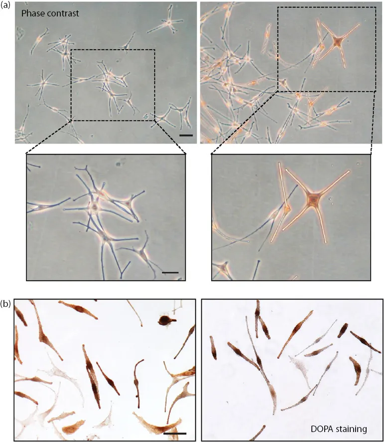
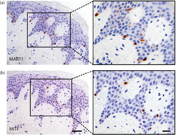
Melanocytes originate from neural crest multipotent precursors and after steps of migration, proliferation, and differentiation finally settle into epidermis and hair follicles as well as extracutaneous sites, for example, mucosa, cardiovascular system, adipose tissue, cochlea, and choroid.7ā€“9 In the skin, they differentiate into dentritic pigment-producing melanocytes (Figure 1.1) and are distributed among keratinocytes of the epidermal basal layer and in hair follicles (Figure 1.2). Synthesized melanin primarily aims at protecting from the harmful effects of UV radiation derived from sunlight as well as, nowadays, from indoor tanning apparatuses, thanks to its ability to absorb UVR and damaging free radicals. The tanning response and the resulting promotion of pigmentation constitute the main protective mechanisms activated following acute and chronic UV exposure by melanocytes and the skin in its entirety.
101367.webp
Figure 1.1 (a) Phase contrast microscopic analysis of primary cultures of normal human melanocytes showing the typical dendritic shape. (b) DOPA staining of human primary melanocytes displaying the cellular brown/black appearance due to the activity of tyrosinase on DOPA substrate. Scale bar: a, b: 50 Āµm.
101403.webp
Figure 1.2 Serial sections of a skin specimen showing the presence of melanocytes identified using the melanocyte markers MART1 (melanoma antigen recognized by T cells 1) (melanosome structural protein) (a), and MITF (microphtalmia-associated transcription factor) (b). Nuclei are counterstained with hematoxylin. Right panels represent higher-magnification images of the black boxed areas. Scale bar: Left panels: 50 Āµm; higher-magnification images on the right panels: 20 Āµm.
Melanocytes actively interact with both epidermal and dermal compartments. Each melanocyte, through its dendrites, is in mutual connection with about 30ā€“40 keratinocytes, constituting the epidermal melanin unit (Figure 1.3), and with dermal fibroblasts, thus establishing a finely balanced network of cellā€“cell crosstalk, ultimately influencing the color of the skin. Differentiated melanocytes display a low growth rate and elevated resistance to apoptosis as a result of their high intrinsic expression of the anti-apoptotic protein Bcl-2.10 Despite variations in the density of melanocytic cells in diverse body areas, their overall number appears constant among human populations. Differences in ethnic color are rather related to the type and quantity of produced melanin and to its transfer, distribution pattern, and degradation into neighboring keratinocytes. There are two main types of melanin synthesized through the multistep process of melanogenesis: red/yellow pheomelanin and brown/black or dark eumelanin, which are both produced in different ratios. In light-skinned people, the predominant melanin type is usually pheomelanin, the melanosomes are smaller and less condensed, and they are transferred to keratinocytes grouped in membrane-bound clusters co...

Table of contents

  1. Cover
  2. Half Title
  3. Title Page
  4. Copyright Page
  5. Contents
  6. Preface
  7. Contributors
  8. 1. Basic concepts on melanocyte biology
  9. 2. Approach to hypopigmentation
  10. 3. Historical review of vitiligo
  11. 4. Epidemiology and classification ofĀ vitiligo
  12. 5. Pathophysiology of vitiligo
  13. 6. Segmental vitiligo
  14. 7. Childhood versus post-childhood vitiligo
  15. 8. Pharmacological therapy of vitiligo
  16. 9. Surgical treatment of vitiligo
  17. 10. Phototherapy and lasers in the treatment of vitiligo
  18. 11. Emerging treatments for vitiligo
  19. 12. Tuberous sclerosis complex
  20. 13. Oculocutaneous albinism
  21. 14. Hermansky-Pudlak syndrome, Chediak-Chigasi syndrome, and Griscelli syndrome
  22. 15. Piebaldism
  23. 16. Waardenburg syndrome
  24. 17. Alezzandrini syndrome, Margolis syndrome, Cross syndrome, and other rare genetic disorders
  25. 18. Mosaic hypopigmentation
  26. 19. Skin disorders causing post-inflammatory hypopigmentation
  27. 20. Infectious and parasitic causes ofĀ hypopigmentation
  28. 21. Melanoma leukoderma
  29. 22. Halo nevi
  30. 23. Drug-induced hypopigmentation
  31. 24. Hypopigmentation from chemical andĀ physical agents
  32. 25. Guttate hypomelanosis and progressive hypomelanosis of the trunk (progressive macular hypomelanosis)
  33. Index