eBook - ePub
Pediatric Emergency Ultrasound
A Concise Guide
- 238 pages
- English
- ePUB (mobile friendly)
- Available on iOS & Android
eBook - ePub
Pediatric Emergency Ultrasound
A Concise Guide
About this book
This concise, portable manual provides practitioners and future practitioners with a basic guide to pediatric emergency ultrasound, enabling them to learn the fundamentals of bedside ultrasound and use these to refresh their skills prior to, or when, performing it on a patient.
Trusted by 375,005 students
Access to over 1.5 million titles for a fair monthly price.
Study more efficiently using our study tools.
Information
Topic
MedicinePART
I
Circulatory system
1Cardiac
2Inferior vena cava
1
Cardiac
Indications
●Aid in identification of cardiac arrest during resuscitation.
●Rapidly assess the global systolic function of a patient presenting with hemodynamic instability or shock.
●Evaluate for evidence of cardiac tamponade in the setting of blunt or penetrating trauma.
●Assess patient’s volume status by evaluation of inferior vena cava.
●Assess patient’s response to resuscitation with serial examinations.
Probe selection
●5-1 MHz low-frequency phased array transducer.
Technique: (Screen indicator located on the left)
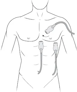
Figure 1.1 Probe placement for focused cardiac ultrasound.
Parasternal short-axis view
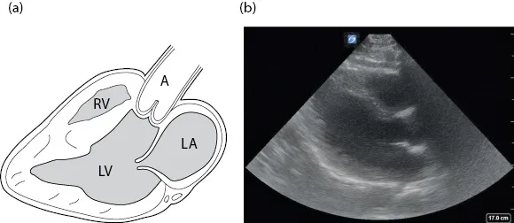
Figure 1.2 Anatomical drawing of parasternal long axis (a) with still image (b).
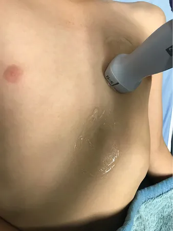
Figure 1.3 Parasternal long view. Probe indicator pointing toward the patient’s left hip.
●Place probe over left parasternal border at the level of the nipple.
●Probe marker should face patient’s right elbow/right hip (90° clockwise from parasternal long-axis view).
Parasternal long-axis view
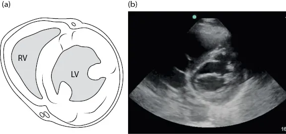
Figure 1.4 Anatomical drawing of parasternal short axis (a) with still image (b).
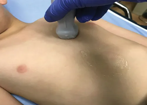
Figure 1.5 Parasternal short view. Probe indicator pointing toward the patient’s right hip.
●Place probe over left parasternal border at the level of the nipple.
●Probe marker should face patient’s left elbow/left hip.
Subxiphoid view
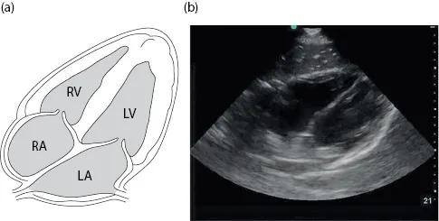
Figure 1.6 Anatomical drawing of subxiphoid view (a) with still image (b).
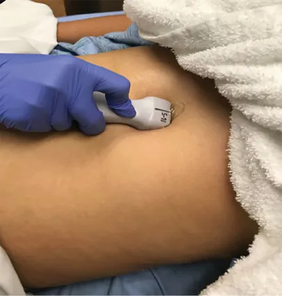
Figure 1.7 Subxiphoid view. Probe fanned from patient’s right to left, using the liver as an acoustic window to visualize the heart.
●Place the probe below the xiphoid process aiming up and into the thoracic cavity.
●Use the liver as an acoustic win...
Table of contents
- Cover
- Half Title
- Title Page
- Copyright Page
- Contents
- Introduction
- Ultrasound probes
- Part I: Circulatory system
- Part II: Respiratory system
- Part III: Musculoskeletal system
- Part IV: Integumentary System
- Part V: Digestive System
- Part VI: Trauma
- Part VII: Renal, urinary, and reproductive systems
- Part VIII: Procedural
- Part IX: Nervous system
- Bibliography
- Index
Frequently asked questions
Yes, you can cancel anytime from the Subscription tab in your account settings on the Perlego website. Your subscription will stay active until the end of your current billing period. Learn how to cancel your subscription
No, books cannot be downloaded as external files, such as PDFs, for use outside of Perlego. However, you can download books within the Perlego app for offline reading on mobile or tablet. Learn how to download books offline
Perlego offers two plans: Essential and Complete
- Essential is ideal for learners and professionals who enjoy exploring a wide range of subjects. Access the Essential Library with 800,000+ trusted titles and best-sellers across business, personal growth, and the humanities. Includes unlimited reading time and Standard Read Aloud voice.
- Complete: Perfect for advanced learners and researchers needing full, unrestricted access. Unlock 1.5M+ books across hundreds of subjects, including academic and specialized titles. The Complete Plan also includes advanced features like Premium Read Aloud and Research Assistant.
We are an online textbook subscription service, where you can get access to an entire online library for less than the price of a single book per month. With over 1.5 million books across 990+ topics, we’ve got you covered! Learn about our mission
Look out for the read-aloud symbol on your next book to see if you can listen to it. The read-aloud tool reads text aloud for you, highlighting the text as it is being read. You can pause it, speed it up and slow it down. Learn more about Read Aloud
Yes! You can use the Perlego app on both iOS and Android devices to read anytime, anywhere — even offline. Perfect for commutes or when you’re on the go.
Please note we cannot support devices running on iOS 13 and Android 7 or earlier. Learn more about using the app
Please note we cannot support devices running on iOS 13 and Android 7 or earlier. Learn more about using the app
Yes, you can access Pediatric Emergency Ultrasound by Marsha A. Elkhunovich, Tarina L. Kang, Marsha Elkhunovich,Tarina Kang,Marsha A. Elkhunovich,Tarina L. Kang in PDF and/or ePUB format, as well as other popular books in Medicine & Emergency Medicine & Critical Care. We have over 1.5 million books available in our catalogue for you to explore.