eBook - ePub
Diagnosis and Management of Soft Tissue Sarcoma
- 272 pages
- English
- ePUB (mobile friendly)
- Available on iOS & Android
eBook - ePub
Diagnosis and Management of Soft Tissue Sarcoma
About this book
Soft tissue sarcoma is an uncommon disease with a historically ominous prognosis, but there has been a great deal of progress in the last two decades in the understanding of these diseases, and accurate prognostic and therapeutic information is now available that removes much of the mystique.
Trusted by 375,005 students
Access to over 1.5 million titles for a fair monthly price.
Study more efficiently using our study tools.
Information
Subtopic
Internal Medicine & Diagnosis1 Historical perspectives and evolution of treatment
Historical perspectives
The term ‘sarcoma’ is derived from the Greek word ‘sarkoma’, meaning a fleshy excrescence. In his writings, Galen (AD 130–200) regarded these fleshy tumors as cancerous and cautioned against surgical intervention.1 With the advent of the light microscope in 1592, a few descriptions were recorded of soft tissue sarcoma, including that of a myxoid lipo-sarcoma by Marcus Severinius (1580–1637)2 and a retroperitoneal liposarcoma by Morgagni (1682–1771).3 The use of thin sections and the achromatic lens, along with other refinements of the microscope, permitted further advances in the recognition of soft tissue sarcoma.
In the eighteenth and nineteenth centuries, Bichat (1771–1801), Abernathy (1780–1848) and Laennec (1781–1826) were among those who made important contributions to the morphologic understanding of cancer. As best we know, the term ‘soft cancer’ was introduced by Wardrop (1782–1869), an Edinburgh surgeon who studied in Vienna, but the name in terms of the definition of soft cancer, as differentiated from carcinoma, has been attributed to Charles Bell (1774–1842), a neuroanatomist. Bell’s book, Surgical Observations, was published in 1816.4
Bichat postulated that anatomic structures consisted of parenchymal cells, which he called ‘filaments’, and stroma, which he called‘fiber’ or ‘tissue’, thus beginning the science of histology. He saw that the stroma was common to many tumors, while the parenchymal cells were specific.5 Abernathy, a pupil of John Hunter, was a prominent surgeon of London who recognized the difference between true neoplasms and non-neoplastic swellings such as cysts, aneurysms, and abscesses. He suggested that tumors be classified by their anatomic structure and offered the first classification of sarcomas.6 Early descriptions to which names have been applied include those of Dupuytren, who in 1832 reported on bladder soft tissue lesions and plantar fibromatosis.
Understanding of the nature of soft tissue sarcoma progressed in the nineteenth century through the studies of cellular pathologists, particularly those of Cruveilhier (1791–1874) and Johannes Muller (1801–1858), who described the cellular origin of various soft tumors. He seems to have coined the term ‘desmoid’ in 1838.4 Many of his ideas were reinforced by Rokitansky (1804–1878). Most importantly, Virchow (1821–1902) advanced the significant view that ‘annis cellula et cellulare’, which literally means ‘where a cell arises, there a cell previously existed’. Sarcomas he defined as ‘new formations of the connective tissue … distinguishable from the corresponding fully evolved tissue by their immaturity’, thereby laying the foundation for the histogenetic classification. His further classification according to microscopic features, which separated sarcomas from carcinomas of epithelial origin, was published in 1863, and is similar to what is accepted today.7 His observations and concepts formed some of the most important milestones in the study of soft tissue sarcomas and paved the way for the development of our current understanding and treatment of these neoplasms.
At that time sarcomas were considered essentially benign, and ‘local growths’ and ‘carcinoma’ were still reserved for lesions that had potential for metastasis. Samuel Gross (1805–1884) described distinctions between sarcoma and carcinoma in the fourth edition of his book, A System of Surgery, in 1866.
When Mallory (1862–1941) introduced his method of staining tissues at the beginning of the twentieth century, the study of soft tissue sarcoma by histopathologic techniques began, and the description and histogenetic classification of sarcomas was advanced by others.
At the Mayo Clinic in the 1920s, Broders suggested that the number of dividing cells in a tumor, mitotic index, might reflect its malignant potential, and gave an illustration of its application in fibrosarcomas.8 The histopathologic grading of sarcomas, vital to the study and treatment of these tumors, was thus begun.
Stout (1885–1967), in a monograph published in 1932, also elucidated on the nature, morphology and treatment of sarcomas.9 His classification of soft tissue sarcoma included the histogenesis, grade of malignancy, including mitotic activity, and cellular as well as stromal organization. Except for minor modifications, this classification remains in use today. The first comprehensive treatise on soft tissue sarcomas10 was the product of his studies.11
Murray and Stout published their classic tissue culture studies of Schwann cells in1942.12 With their later studies of synovial tissue and synovial sarcoma, they confirmed the mesenchymal origin of synovial sarcomas, even though these tumors may show epithelial characteristics.13
Classic contributions to the description and histogenetic classifications of sarcoma were made at the Memorial Hospital for Cancer and Allied Diseases, starting with Dr James Ewing (1866–1943) (Figure 1.1). James Ewing was the first Professor of Pathology at Cornell University Medical Center. Having graduated in 1891 with his MD degree, he assumed the position of Chief of Pathology at Memorial Hospital in 1899 at the age of 33. When he published the first edition of his monograph, Neoplastic Diseases, in 1919, his observations and concepts of tumors laid the foundation for the surgical pathology of neoplasms.14 He completed the book despite trigeminal neuralgia. In 1926 neurosurgery by Dr Harvey Cushing replaced the paroxysms of tic doloureux with a painful anesthesia which disabled him for the rest of his life.
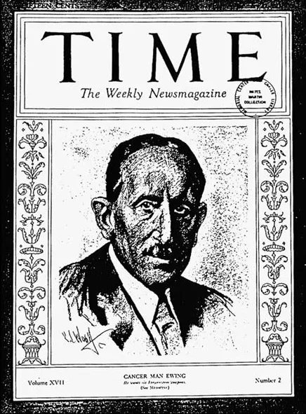
Figure 1.1Dr James Ewing.
In a succession of editions, he gave a clear classification of soft tissue sarcoma and stated that ‘Sarcoma is a malignant tumor composed of cells of the connective tissue type … This definition is based on the morphology of the tumor cells and on their histogenesis.’ He listed benign and malignant counterparts of tumors arising from fibrous tissue, cartilage, bone, myxomatous tissue, fatty tissue, blood and lymphatic vessels, smooth and striated muscle, and vascular endothelium. He also recognized that the accepted scope of sarcomas has been subject to much revision, since there is often much difficulty in determining the origin of cellular tumors. One of his most important Ewing’s sarcoma, first described in 1920.15 The suggestion that grade was of importance in the outcome of sarcomas was first emphasized in the fourth edition of Neoplastic Diseases, published in 1931. 4
William Coley (Figure 1.2) in 1889 had treated the 17-year-old Elizabeth Dashiell at Memorial Hospital for Cancer and Allied Diseases for an extremity sarcoma. Despite his surgical efforts, this young friend of John D Rockefeller Jr died from her disease in June 1890. This had a significant effect on Coley and influenced him to study sarcoma. He continued to study the treatment and outcome of patients of his mentor, William Tillinghast Bull. In the lower east side of Manhattan, Coley found a patient with recurrent sarcoma. After the patient had multiple recurrences resected from his neck, he was then surprisingly cured by a postoperative erysipelas infection. Based on this, Coley started using Coley’s toxins in 1892 and instituted the advent of immunotherapy in cancer.
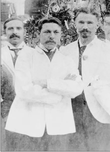
Figure 1.2 Dr William Coley.
A great many contributions to pathologic evaluation have come from Memorial Sloan-Kettering Cancer Center, from Ewing to Stout in 1942, who introduced the term ‘hemangio-pericytoma’. Moreover, Stout’s classification of liposarcomas in 1944 was a first, as was his description, with Ackerman, of leiomyo-sarcoma of soft tissue in 1947. He published a comprehensive listing of tumors of soft tissue in an Armed Forces Institute of Pathology fasicle in 1953.10
In 1948, Stewart and Treves at Memorial Hospital (Figures 1.3 and 1.4) described lymphangiosarcoma, a highly malignant tumor, in post-mastectomy patients with chronic lymphedema of the upper limb.16 Since their original description, lymphangiosarcoma has been found in non-edematous post-mastectomy upper limbs17 and in patients with limbs congenitally lymphadematous or edematous due to filariasis or trauma.18 Stewart, in 1952, reported the first case of alveolar soft part sarcoma.
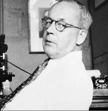
Figure 1.3 Dr Frederick Stewart.
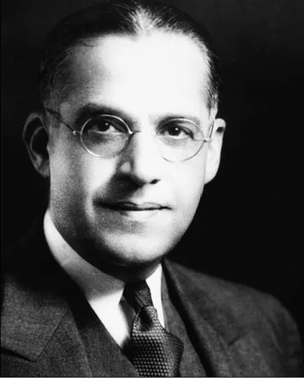
Figure 1.4 Dr Norman Treves.
In the 1940s to1960s, the term ‘giant and spindle cell sarcoma’ was often used as a descriptive pathologic diagnosis for many soft tissue sarcomas. Myxoid and histiocytic cellular elements often combined with a varying amount of collagenous tissue in the stroma in these neoplasms. In 1967, Stout and Lattes described the morphology of these tumors and coined the term (malignant) fibrous histiocytoma, which they considered to be embryonal forms of fibroblastic n...
Table of contents
- Cover page
- Title page
- Copyright Page
- Introduction
- Acknowledgements
- 1: Historical perspectives and evolution of treatment
- 2: Incidence, etiology
- 3: Pathologic classification
- 4: Clinical features, diagnosis and extent of disease evaluation
- 5: Staging
- 6: Prognostic factors
- 7: Clinical and pathologic correlates
- 8: Principles of management
- 9: Adjuvant management
- 10: Special sites
- 11: Special techniques
- 12: Treatment of recurrence
- 13: Future directions
Frequently asked questions
Yes, you can cancel anytime from the Subscription tab in your account settings on the Perlego website. Your subscription will stay active until the end of your current billing period. Learn how to cancel your subscription
No, books cannot be downloaded as external files, such as PDFs, for use outside of Perlego. However, you can download books within the Perlego app for offline reading on mobile or tablet. Learn how to download books offline
Perlego offers two plans: Essential and Complete
- Essential is ideal for learners and professionals who enjoy exploring a wide range of subjects. Access the Essential Library with 800,000+ trusted titles and best-sellers across business, personal growth, and the humanities. Includes unlimited reading time and Standard Read Aloud voice.
- Complete: Perfect for advanced learners and researchers needing full, unrestricted access. Unlock 1.5M+ books across hundreds of subjects, including academic and specialized titles. The Complete Plan also includes advanced features like Premium Read Aloud and Research Assistant.
We are an online textbook subscription service, where you can get access to an entire online library for less than the price of a single book per month. With over 1.5 million books across 990+ topics, we’ve got you covered! Learn about our mission
Look out for the read-aloud symbol on your next book to see if you can listen to it. The read-aloud tool reads text aloud for you, highlighting the text as it is being read. You can pause it, speed it up and slow it down. Learn more about Read Aloud
Yes! You can use the Perlego app on both iOS and Android devices to read anytime, anywhere — even offline. Perfect for commutes or when you’re on the go.
Please note we cannot support devices running on iOS 13 and Android 7 or earlier. Learn more about using the app
Please note we cannot support devices running on iOS 13 and Android 7 or earlier. Learn more about using the app
Yes, you can access Diagnosis and Management of Soft Tissue Sarcoma by Murray Brennan, Jonathan Lewis, Murray Brennan,Jonathan Lewis in PDF and/or ePUB format. We have over 1.5 million books available in our catalogue for you to explore.