eBook - ePub
An Atlas of Three- and Four-Dimensional Sonography in Obstetrics and Gynecology
- 216 pages
- English
- ePUB (mobile friendly)
- Available on iOS & Android
eBook - ePub
An Atlas of Three- and Four-Dimensional Sonography in Obstetrics and Gynecology
About this book
The use of three-dimensional ultrasound has expanded, particularly in obstetrics and gynecology clinical practice, necessitating a reference covering this and other emerging technologies. This volume presents ultrasound images in full colour accompanied by extensive captions and expert textual commentary. It provides authoritative coverage of this important technology that can help diagnose a host of conditions that heretofore would go unseen or require invasive surgery to diagnose.
Trusted by 375,005 students
Access to over 1.5 million titles for a fair monthly price.
Study more efficiently using our study tools.
Information
Topic
Medicine1: Introduction to three-and four-dimensional ultrasound
K.Baba
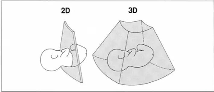
Figure 1 Two-dimensional data for conventional 2D ultrasound (left) and 3D data for 3D ultrasound (right)
INTRODUCTION
A conventional ultrasound scanner acquires two-dimensional (2D) information and displays a sectional image only. A three-dimensional (3D) ultrasound scanner acquires three-dimensional information (Figure 1), generates and displays various kinds of sectional and 3D images from the acquired 3D data. A 3D ultrasound scanner in common use generates images with a computer. Imaging technology has developed rapidly since a 3D image of a live fetus in utero was first demonstrated by Baba and colleagues in 1986 by using their original computerized 3D ultrasound system1. The system could also handle Doppler flow data and demonstrated placental blood flows2.
HOW A THREE-DIMENSIONAL ULTRASOUND SCANNER WORKS
Various images are obtained through the following processes in 3D ultrasound:
- Acquisition of 3D data (3D scanning);
- Construction of a 3D data set;
- Volume visualization.
Acquisition of three-dimensional data
Three-dimensional data are usually acquired in the form of a large number of consecutive tomographic images through movements of a conventional 2D ultrasound probe. Three 3D scanning methods are shown in Figure 2. Each tomographic image should be acquired with its positional information for the subsequent processing. Accurate positional information about the probe can be obtained through an electromagnetic position sensor attached to the probe (Figure 3). However, 3D probes are widely used because of their ease in use. Each 3D probe has a built-in 2D ultrasound probe, which tilts in the 3D probe and thus 3D data are acquired automatically (Figure 4).
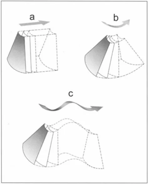
Figure 2 3D scanning methods. (a) Parallel scanning; (b) fan-like scanning; (c) free surface scanning3
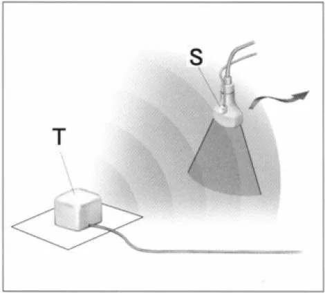
Figure 3 A position sensor attached to a probe detects a relative position of the probe. T, transmitter; S, electromagnetic sensor12
Construction of a three-dimensional data set
The tomographic images obtained through 3D scanning must be constructed three-dimensionally into a 3D data set for further computer processing (Figure 5). This construction process involves interpolation and filtering to improve the data quality3. A 3D data set is composed of a set of voxels (volume elements). Each voxel has a gray value.
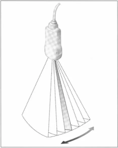
Figure 4 3D scanning by a 3D probe12
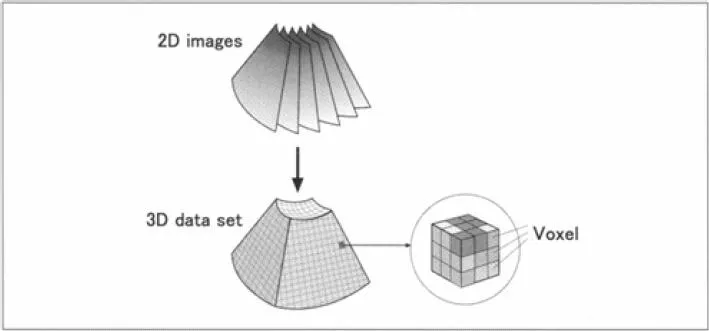
Figure 5 Construction of a 3D data set12
Volume visualization
A 3D data set should be processed by a computer so that it can be displayed on a 2D screen. This process is called volume visualization. Three methods have been commonly used for volume visualization in 3D ultrasound:
- Section reconstruction;
- Volume rendering;
- Surface rendering.
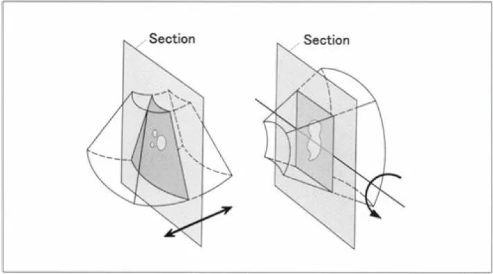
Figure 6 Arbitrary section display12
Section reconstruction
A sectional image can be obtained by cutting a 3D data set. An arbitrary section can be selected and displayed through translation and rotation of the 3D data set (Figure 6). Three orthogonal sections are usually displayed on a screen immediately after 3D scanning (Figure 7), and then either standardized or suitable sections of the object are displayed through translation and rotation (Figure 8). Although displayed images by section reconstruction are sectional images, they are sometimes very useful for diagnosis, because some images cannot be obtained by conventional 2D ultrasound. Three orthogonal sections may also be arranged three-dimensionally (Figure 9).
Volume rendering
Various kinds of 3D images can be generated by volume rendering. A viewing direction is first determined and a smaller 3D data set for rendering (3D image generation) is extracted from the original 3D data set to eliminate unnecessary parts around the object as much as possible (Figure 10). Both the setting of the viewing direction and extraction of the smaller 3D data set for rendering can be performed on three orthogonal planes by translation and rotation of the 3D data set and by setting a region of interest (ROI) (Figure 11).
A 3D data set for rendering is projected directly on a projection plane in volume rendering (Figure 12). Rays are assumed from each pixel on the projection plane into the 3D data set. The brightness of each pixel is determined based on the gray values of voxels on each corresponding ray.
The fetal skeleton can be seen when only the maximal gray value on each ray is displayed on the projection plane (Figure 13). A 3D image of cystic parts is obtained when only the minimal gray value on each ray is displayed on the projection plane (Figure 14). A transparent 3D image, just like an X-ray photograph, is obtained by displaying the average gray value on each ray (Figure 15).
A fetal surface image (Figure 16) can also be obtaine...
Table of contents
- Cover Page
- Title page
- Copyright page
- List of principal contributors
- Foreword
- 1: Introduction to three-and four-dimensional ultrasound
- 2: Three-and four-dimensional ultrasound in human reproduction
- 3: Three-dimensional ultrasound and three-dimensional power Doppler in the assessment of adnexal masses
- 4: Three-and four-dimensional visualization in early pregnancy
- 5: Three-and four-dimensional images of fetal malformations
- 6: Clinical application of four-dimensional sonography in perinatal medicine
- 7: Fetal echocardiography for the general practitioner: how to use three-and four-dimensional imaging to obtain a cardiac screening index
- 8: Three-and four-dimensional sonography in multiple pregnancy
- 9: Three-dimensional ultrasound in the assessment of the neonatal brain
Frequently asked questions
Yes, you can cancel anytime from the Subscription tab in your account settings on the Perlego website. Your subscription will stay active until the end of your current billing period. Learn how to cancel your subscription
No, books cannot be downloaded as external files, such as PDFs, for use outside of Perlego. However, you can download books within the Perlego app for offline reading on mobile or tablet. Learn how to download books offline
Perlego offers two plans: Essential and Complete
- Essential is ideal for learners and professionals who enjoy exploring a wide range of subjects. Access the Essential Library with 800,000+ trusted titles and best-sellers across business, personal growth, and the humanities. Includes unlimited reading time and Standard Read Aloud voice.
- Complete: Perfect for advanced learners and researchers needing full, unrestricted access. Unlock 1.5M+ books across hundreds of subjects, including academic and specialized titles. The Complete Plan also includes advanced features like Premium Read Aloud and Research Assistant.
We are an online textbook subscription service, where you can get access to an entire online library for less than the price of a single book per month. With over 1.5 million books across 990+ topics, we’ve got you covered! Learn about our mission
Look out for the read-aloud symbol on your next book to see if you can listen to it. The read-aloud tool reads text aloud for you, highlighting the text as it is being read. You can pause it, speed it up and slow it down. Learn more about Read Aloud
Yes! You can use the Perlego app on both iOS and Android devices to read anytime, anywhere — even offline. Perfect for commutes or when you’re on the go.
Please note we cannot support devices running on iOS 13 and Android 7 or earlier. Learn more about using the app
Please note we cannot support devices running on iOS 13 and Android 7 or earlier. Learn more about using the app
Yes, you can access An Atlas of Three- and Four-Dimensional Sonography in Obstetrics and Gynecology by David Jackson, Asim Kurjak, David Jackson,Asim Kurjak in PDF and/or ePUB format, as well as other popular books in Medicine & Gynecology, Obstetrics & Midwifery. We have over 1.5 million books available in our catalogue for you to explore.