Chapter 1
The human body and principles of homeostasis
Introduction
This book is about anatomy and physiology; the two branches of science that will help you understand the human body. Identifiable within these sciences is the central concept referred to as homeostasis.
Definitions
- Human anatomy refers to the study of the structure of the body.
- Physiology is concerned with the mechanisms of human bodily function.
- Homeostasis refers to the automatic, self-regulating processes necessary to maintain the ‘normal’ state of the body’s environment despite changes in the environment outside the body.
Collectively, anatomical structure, physiological function and the maintenance of homeostasis enable the body to attain the basic needs necessary for health and a ‘normal’ life. Before contemplating the principles of homeostasis, the reader should become familiar with how the body is organized.
Anatomical organization
The outside of the human body has a definite and recognizable shape; the inside contains the organs, which are specifically located relative to each other. The anatomical position of the body provides a reference point when studying or describing the position of body structures. This position is when the person stands erect, facing forward, with arms at his/her side with palms facing forwards. For example, organs such as the heart are drawn according to this convention, so those features on the left side appear on the right, almost as though the observer is looking into someone’s chest.
Box 1.1 Application: positioning the patient on the operating table
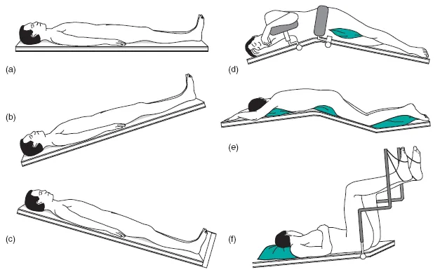
The position of the patient on the operating table usually depends on the operation being carried out and the surgeon’s preference. The needs of the anaesthetist are also considered, since the position must enable him/her to maintain anaesthesia in complete safety. However, the patient may have a predisposing condition, which prevents the preferred position from being used and an alternative or revised position must be sought. It is important for the perioperative practitioner to have an in-depth knowledge of the various table positions (Figure 1.1 see p.) and the equipment required for each case. Particular attention is applied to patient safety; the operating table is very narrow and the patient must be placed securely on the table. The use of pressure relieving devices is essential as patients can remain in one position for several hours, and ischaemic changes to areas exposed to pressure can cause pressure areas/sores, potentially prolonging the patient’s hospital stay. Over-extension of joints and limbs must be avoided; poor positioning can lead to nerve and muscle damage and dislocation of joints. Any exposed areas of the body, which may come into contact with metal (table or attachments), must be protected to avoid inadvertent diathermy burns. Some positions might have special requirements, making them complex and time consuming to achieve, possibly involving a number of personnel.
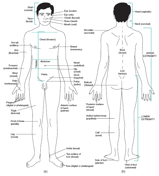
A set of standard anatomical terms is used to describe each part of the body, the position of body structures and their geographical relationships with each other. Many of these regional terms are illustrated in Figure 1.2. The main terms used relate to ‘planes of the body’, ‘relative positioning of organs’ and ‘body cavity’ (see below, see p.).
Planes of the body
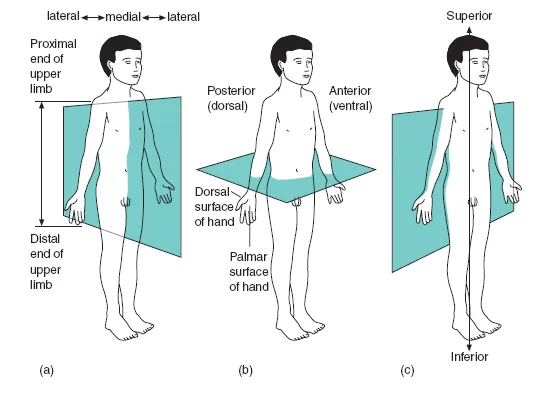
Body structures are described by the perioperative practitioner in relation to three planes or imaginary lines which run through the body. These are called median (or mid-sagittal), transverse (or horizontal) and coronal (or frontal) lines (Figure 1.3). The median plane passes directly along the mid-line of the body, dividing the body into perfectly symmetrical right and left halves. The transverse plane passes horizontally through the body dividing it into upper and lower portions. The coronal plane divides the body into front and back portions.
Terms of relative position
The perioperative practitioner needs to understand the position of structures relative to each other and also to use directional terms (see Figure 1.3). For example: anterior and ventral are used interchangeably and refer to the front surface of the body. They also have a broader meaning, in the sense of a structure being closer to the front of the body. As an example: ‘anterior abdominal wall’ or ‘the heart is anterior to the spine’. In the same way, posterior may refer to the back surface of the body or it may mean a structure is nearer to the back of the body. For example, the oesophagus (food pipe) is posterior to the heart. ‘Dorsal’ is similarly linked to ‘posterior’. Likewise medial indicates that a structure is towards the mid-line of the body, whereas lateral designates that a structure is away from the mid-line of the body. ‘Proximal’ indicates that the structure concerned is nearer the attached end of a limb or thus the trunk of the body, whilst a ‘distal’ structure is farther away from the attached end of a limb and the trunk. For example, the humerus is proximal to the radius, whereas the phalanges are distal to the carpals.
EXERCISES
1 Review the common surgical procedures employed when the patient is placed in the positions identified in Figure 1.1a–f and identify the equipment required for each surgical procedure cited.
2 Match the pressure areas indicated by Lists A, B and C – with the following positions
a The supine position is associated with the pressure areas identified in List ____
b The lateral position is associated with the pressure areas identified in List ____
c The prone position is associated with the pressure areas identified in List ____
List A
Medial and lateral
Malleolus
Greater trochanter
Ilium
Ribs
Ear
Acromion process
List B
Toes
Patella
Male genitalia
Breast
Acromion process
Cheek
Ear
List C
Calcaneus
Sacrum and coccyx
Thoracic vertebrae
Scapulae
Occiput
See the end of this chapter for the answers to question 2.
EXERCISES
Look up in a biological dictionary what the following terms mean: afferent, efferent, peripheral, deep, superficial, internal and external.
Identify the location of the following bones: humerus, radius, phalanges and carpals.
Figure 1.1 Common positioning of the patient for various surgical procedures:
a the supine position,
b the Trendelenburg position,
c the reverse Trendelenburg position,
d the lateral position,
e the prone position,
f the lithotomy position.
Perioperative practitioners must be familiar with the terminology used in theatre, speaking the same language, to avoid mistakes. Early in the practitioner’s career, the anatomical terminology may be unfamiliar and difficult to understand. But once the practitioner has mastered an understanding of basic words, including prefixes (the beginning of a word), roots (the main body of the word) and suffixes (the ends of words) you will be able to apply these principles to derive the meaning of terminology. For example, once you understand the prefix: ‘electro-’, means electrical, the root ‘cardiac’ refers to the heart and the suffix ‘-gram’ means recording, then the meaning of the term ‘electrocardiogram’ becomes apparent.
Figure 1.2 Anatomical position
The common names and anatomical terms (indicated in brackets) for many of the regions of the human body.
a anterior view,
b posterior view.
Figure 1.3 Planes through the body:
a medial (midsagittal) plane,
b transverse plane,
c coronal (frontal) plane.
Q. Which plane divides the body into:
a anterior and posterior parts?
b right and left parts which are mirror images of each other?
c superior and inferior portions?
Q. Is the hip proximal or distal to the knee?
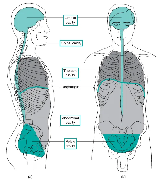
Body cavities
The body can be divided into cavities produced by the bony skeleton (Figure 1.4), and these contain the internal organs (or viscera). The main body cavities are:
- the cranial cavity. The bones of the skull enclose this. This cavity accommodates and protects the brain.
- the spinal cavity. This is formed by a hole (foramen = ‘window’) running through the vertebral column. This is called the vertebral canal and it accommodates and protects the spinal cord.
- the thoracic cavity. This is the upper cavity of the trunk of the body. Its confines are the breastbone (or sternum), ribs and associated intercostal muscles (inter- = between, -cost = rib) and cartilage, vertebral column, diaphragm and the structures below the neck. The cavity contains the windpipe (or trachea), two lungs, the heart and its great vessels, the food pipe (or oesophagus) and associated nerves and lymphatic supply. The space between the lungs, occupied by the heart, is called the mediastinum.
- the abdominal cavity. This is the large lower portion of the trunk. The diaphragm, the pelvic cavity, the spine, abdominal muscles and lower ribs confine it. It accommodates the organs concerned with digestion and absorption of nutrients, and other organs associated with these functions. These are the gall bladder, liver, spleen and pancreas. In addition the kidneys, ureters (superior regions) and adrenal glands are also located in this region.
- the pelvic cavity. This is the lowest portion of the trunk, a continuation of the abdominal cavity. Its boundaries are the bony pelvis, sacrum and muscles of the pelvic floor. In the female it contains all the reproductive structures; in the male, however, only some reproductive structures are contained. The cavity of both sexes contains the ureters (lower regions), bladder and urethra. Other pelvic organs are part of the small intestine and the last part of the colon, the rectum and anal canal. It also includes the openings for the urethra, vagina (in the female) and anus.
Blood vessels, lymphatic nodes and associated nerves are located in all the cavities.
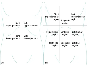
Sometimes it is necessary to be more precise in locating organs within the body. Thus the body is further subdivided into quadrants and the nine regions associated with the abdomen and pelvic areas (see Figure 1.5). This level of precision is required to determine the sites for surgical incision.
Figure 1.4 Lateral and anterior views of the human body showing major cavities:
a right lateral view,
b anterior view.
Q. What are the confines of the following cavities:
- thoracic cavity,
- abdominal cavity,
- pelvic cavity.
Figure 1.5 Anatomical sub-divisions of the abdomen and pelvic regions:
a quadrants of the abdomen,
b the nine regions of the abdominal and pelvic regions.
Q. Identify the quadrant that confines the stomach.
Q...