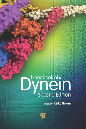
Handbook of Dynein (Second Edition)
eBook - ePub

Handbook of Dynein (Second Edition)

Keiko Hirose, Keiko Hirose

  1. 420 pages
  2. English
  3. ePUB (mobile friendly)
  4. Available on iOS & Android
eBook - ePub

Handbook of Dynein (Second Edition)

Keiko Hirose, Keiko Hirose

Book details
Book preview
Table of contents
Citations

About This Book

Dyneins are molecular motors that are involved in various cellular processes, such as cilia and flagella motility, vesicular transport, and mitosis. Since the first edition of this book was published in 2012, there has been a significant breakthrough: the crystal structures of the motor domains of cytoplasmic dynein have been solved and the previously unknown details of this huge and complex molecule have been unveiled. This new edition contains 14 chapters written by researchers in the US, Europe, and Asia, including 3 new chapters that incorporate new fields. The other chapters have also been substantially updated. Compared with the earlier edition, this book focuses more on the motile mechanisms of dynein, especially by biophysical methods such as cryo-EM, X-ray crystallography, and single-molecule nanometry. It is a major handbook for frontline researchers as well as for advanced students studying cell biology, molecular biology, biochemistry, biophysics, and structural biology.

Frequently asked questions

How do I cancel my subscription?
Simply head over to the account section in settings and click on “Cancel Subscription” - it’s as simple as that. After you cancel, your membership will stay active for the remainder of the time you’ve paid for. Learn more here.
Can/how do I download books?
At the moment all of our mobile-responsive ePub books are available to download via the app. Most of our PDFs are also available to download and we're working on making the final remaining ones downloadable now. Learn more here.
What is the difference between the pricing plans?
Both plans give you full access to the library and all of Perlego’s features. The only differences are the price and subscription period: With the annual plan you’ll save around 30% compared to 12 months on the monthly plan.
What is Perlego?
We are an online textbook subscription service, where you can get access to an entire online library for less than the price of a single book per month. With over 1 million books across 1000+ topics, we’ve got you covered! Learn more here.
Do you support text-to-speech?
Look out for the read-aloud symbol on your next book to see if you can listen to it. The read-aloud tool reads text aloud for you, highlighting the text as it is being read. You can pause it, speed it up and slow it down. Learn more here.
Is Handbook of Dynein (Second Edition) an online PDF/ePUB?
Yes, you can access Handbook of Dynein (Second Edition) by Keiko Hirose, Keiko Hirose in PDF and/or ePUB format, as well as other popular books in Biological Sciences & Biology. We have over one million books available in our catalogue for you to explore.

Information

Year
2019
ISBN
9780429664717
Edition
2

Chapter 1


Dyneins: Ancient Protein Complexes Gradually Reveal Their Secrets

Linda A. Amosa and Keiko Hiroseb
aMRC Laboratory of Molecular Biology, Cambridge CB2 0QH, UK
bBiomedical Research Institute, National Institute of Advanced Industrial Science and Technology (AIST), Tsukuba, Ibaraki 350-8565, Japan
The history of research on dynein began with EM images showing dynein ‘arms’ forming crossbridges between the doublet microtubules in a ciliary axoneme. In the half-century since then, our view of these amazing macromolecules has gradually reached a point where some of its protein domains including the dynein motor domain have been seen at atomic resolution. Also, the origin and evolution of the various dynein components are now becoming clearer.

1.1 Introduction

Dynein was first identified and named by Ian Gibbons in the 1960s as an ATPase that could be extracted from cilia and flagella. The number of papers written per year has increased steadily ever since (Fig. 1.1). The complex structural and functional secrets of this microtubule (MT) motor were very gradually unlocked. In Fig. 1.2 we summarize some of the important advances that were made in the past half-decade. Recently, however, progress has accelerated greatly, thanks to a variety of tools that were not available originally, such as sequencing of whole genomes, success in producing recombinant dynein, X-ray crystallography, EM cryo-microscopy and tomography, and single-molecule measurements. This makes it difficult to summarize all important new contributions. Current understanding of dynein’s structure and motile mechanism, and its wide range of roles in vivo, are described in more detail in subsequent chapters.
images
Figure 1.1 Increasing number of published papers on dynein.

1.2 Dynein Molecular Structure Coming into View

1.2.1 Axonemal Arms

Dynein was first seen in an electron microscope (EM) as two rows of ‘arms’ on each doublet MT in thin sections of flagella (see Chapter 11, Chapter 12 and Chapter 13) whose fine structure had been preserved with a new chemical fixative, glutaraldehyde [1]. The image in Fig. 1.3i is an example of similar results obtained by Gibbons and Grimstone [39], who improved the contrast in their sections by introducing a novel staining method. A few years later, a protein having ATPase activity was extracted from Tetrahymena cilia and named ‘dynein’ by Gibbons and Rowe [41]. A fraction that was characterized by ultracentrifugation as 14S molecules was seen by EM (Fig. 1.3ii) as individual globular particles; another fraction, consisting of larger complexes appeared to be a longish linear polymer (30S dynein), whose identity is still a little mysterious. A decade later, outer arm of Tetrahymena cilia, before and after extraction from axonemes, appeared in negative stain as a linear complex of three subunits [160] (Fig. 1.3iii). It became also clear that outer arms are arranged with a periodicity of ∼24 nm in axonemes.
images

images
Figure 1.2 Dynein discovery timeline. Abbreviations: EM, electron microscopy; MT, microtubule; HC, heavy chain; IC, intermediate chain; LC, light chain; ODA, outer dynein arm; IDA, inner dynein arm; OAD, outer-arm dynein (proteins); CD, cytoplasmic dynein; IFT, intraflagellar transport; MTBD, microtubule-binding domain; EB1, end-binding protein. ‘arms’ are the projections seen extending from doublet MTs (see Fig. 1.3i).
Conformational changes were observed between the dynein arms crossbridging two doublet MTs and those unbound to the B-tubule [159] (Fig. 1.3iv), and between the arms in the presence and absence of ATP [143]. It was found that the binding of arms to the B-tubule is nucleotide-dependent, but there was controversy as to whether the arms dissociate from the B-tubule with ATP. In 1982, Goodenough and Heuser [46] first saw a thin stalk extending from the globular dynein head, in axonemes that had been rapidly frozen to preserve their structure (Fig. 1.3v–viii). Conformational changes in the presence/absence of ATP were also clearly demonstrated [46, 126] (Fig. 1.3v,vii). At this point, therefore, the main structural features of dynein molecules (Chapter 2, Chapter 3 and 4, and 13) had already been observed but they could not properly be understood until the HC sequence had been determined in 1991 [38, 101], which paved the way both for identification of the MT-binding region in 1997 [32, 75] and recognition of dynein as a member of the superfamily of AAA+ proteins in 1999 [98].
images
Figure 1.3 Electron micrographs of axonemal dynein arms, showing the gradual increase in resolution and understanding of the structure, (i) TEM image of a stained thin section cut through glutaraldehyde-fixed plastic-embedded flagella [39]. (ii) Top-left: ‘30S dynein’ polymers extracted from Tetrahymena axonemes and shadowed with platinum; bottom-left: 30S dynein image after translational averaging with 14 nm steps; right: separate ‘14S dynein’ subunits viewed in negative stain [41]. (iii) Negatively stained Tetrahymena dynein arms arranged on a doublet MT (bottom) and extracted ‘14S dynein’ subunits (top), both showing three subunits [160]. (iv) Tetrahymena doublet MTs crossbridged with outer arms, in two conformational states [159]. (v–vii) Chlamydomonas (v–vi) and sea urchin (vii) axonemes, demembranated and quick-frozen before being shadowed with platinum [46, 126]. In v and vii, the side views of axonemes fixed in the ATP-free solution to trap the dynein arms in the rigor state (top) and those relaxed by incubation in vanadate plus ATP (bottom) show markedly different conformations of dynein arms. The stalks connecting the dynein arms to the B-tubule of the next doublet-MT are clearly observed in both states. The basal end is on the right. In vi, an axoneme is viewed from tip to base and the arrow indicates a relaxed dynein outer arm. (viii) View of a doublet MT from inside an axoneme. The radial spokes (S), in groups of three, are seen end-on and, next to them, the dynein inner arms form two pairs (D) of distinct individual heads, then a triad (T) [47], which correspond to 7 different dynein species [63]. (ix–xiii) 3D tomographic images of quick-frozen axonemes. ix is a view similar to viii, showing inner dynein arms [13] (see Chapter 13). (x) A doublet MT with outer and inner arms, viewed from tip to base [52]. (xi) Native Chlamydomonas outer arms [58, 99]; the two images show very similar details, each arm being a stack of three dy...

Table of contents

  1. Cover
  2. Half Title
  3. Title Page
  4. Copyright Page
  5. Table of Contents
  6. Preface
  7. 1 Dyneins: Ancient Protein Complexes Gradually Reveal Their Secrets
  8. 2 Structural and Functional Analysis of the Dynein Motor Domain
  9. 3 Electron Microscopy Studies of Dynein: From Subdomains to Microtubule-Bound Assemblies
  10. 4 Subunit Architecture of the Cytoplasmic Dynein Tail
  11. 5 Measuring the Motile Properties of Single Dynein Molecules
  12. 6 Mechanics of Dynein Motility
  13. 7 Interactions of Multiple Dynein Motors Studied Using DNA Scaffolding
  14. 8 Cytoplasmic Dynein Force Regulation in vitro and in vivo
  15. 9 Dynein in Endosome and Phagosome Maturation
  16. 10 Dynein in Intraflagellar Transport
  17. 11 Diversity of Chlamydomonas Axonemal Dyneins
  18. 12 Motility of Axonemal Dyneins
  19. 13 Axonemal Dyneins in Cilia and Flagella
  20. 14 Regulatory Mechanism of Axonemal Dynein
  21. Index
Citation styles for Handbook of Dynein (Second Edition)

APA 6 Citation

[author missing]. (2019). Handbook of Dynein (Second Edition) (2nd ed.). Jenny Stanford Publishing. Retrieved from https://www.perlego.com/book/1604558/handbook-of-dynein-second-edition-pdf (Original work published 2019)

Chicago Citation

[author missing]. (2019) 2019. Handbook of Dynein (Second Edition). 2nd ed. Jenny Stanford Publishing. https://www.perlego.com/book/1604558/handbook-of-dynein-second-edition-pdf.

Harvard Citation

[author missing] (2019) Handbook of Dynein (Second Edition). 2nd edn. Jenny Stanford Publishing. Available at: https://www.perlego.com/book/1604558/handbook-of-dynein-second-edition-pdf (Accessed: 14 October 2022).

MLA 7 Citation

[author missing]. Handbook of Dynein (Second Edition). 2nd ed. Jenny Stanford Publishing, 2019. Web. 14 Oct. 2022.