This is a test
- 160 pages
- English
- ePUB (mobile friendly)
- Available on iOS & Android
eBook - ePub
Book details
Book preview
Table of contents
Citations
About This Book
The Physiological Basis of Behaviour deals with the basic structures of the central nervous system, the techniques used in neuroscience and examnines how drugs affect the brain.
Frequently asked questions
At the moment all of our mobile-responsive ePub books are available to download via the app. Most of our PDFs are also available to download and we're working on making the final remaining ones downloadable now. Learn more here.
Both plans give you full access to the library and all of Perlego’s features. The only differences are the price and subscription period: With the annual plan you’ll save around 30% compared to 12 months on the monthly plan.
We are an online textbook subscription service, where you can get access to an entire online library for less than the price of a single book per month. With over 1 million books across 1000+ topics, we’ve got you covered! Learn more here.
Look out for the read-aloud symbol on your next book to see if you can listen to it. The read-aloud tool reads text aloud for you, highlighting the text as it is being read. You can pause it, speed it up and slow it down. Learn more here.
Yes, you can access The Physiological Basis of Behaviour by Kevin Silber in PDF and/or ePUB format, as well as other popular books in Psychology & History & Theory in Psychology. We have over one million books available in our catalogue for you to explore.
Information
1
Methods of investigating brain function
Introduction
Gaining knowledge about the workings of the brain is not an easy task. In the early part of the twentieth century, neuroanatomists relied on a keen eye and a little help from the light microscope. However, during this century huge technological advances have improved the methodologies available to the neuroscientist. In this chapter we will explore some of these methodologies and evaluate their contribution to our knowledge.
The chapter is divided into two parts: neuroanatomical techniques, which allow us to learn more about the structure of the brain; and neurophysiological techniques, which allow us to investigate the functioning of the brain.
Neuroanatomical techniques
There are a number of different techniques which allow neuroanatomists to investigate the structure of the brain. These range from simple observations using microscopes, to chemical staining methods allowing us to pinpoint nerve cells and fibre pathways, to brain scanning techniques that allow us to construct 3D images of the whole brain.
Electron microscopy
Rationale
This method allows us to see in detail the size and shape of neurons and their components (Figure 1.1).
Description
The electron microscope uses electrons instead of light to magnify the image. This allows us to see actual synapses which are only a few ten-millionths of a millimetre in diameter. (The unit which represents one ten-millionth of a millimetre is the angstrom which has the symbol Å.) Another type of electron microscope is the scanning electron microscope, which lets us see the image in 3D, although the magnification has to be very slightly reduced.
Electron microscopy is used routinely today to examine changes in the structure of neurons either after brain damage or in response to information processing. For example, Spinelli et al. (1980) showed that the structure of neurons in a sensory area of a kitten’s brain changed after it had learned a simple conditioning task.
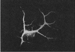
Figure 1.1 An electron photomicrograph of a neuron
Evaluation
This method gives us a very clear picture of the minute detail of neurons. However, the equipment is extremely expensive and the method can only be used post mortem.
Chemical staining
Rationale
Staining methods allow us to trace the extent of features of the nervous system. For example, we can examine where cell bodies are or search for where the neurons from a particular region of the brain project to. There are a vast number of different stains that can be applied to brain tissue sections and each has a different feature that it highlights best.
Description
To stain brain tissue a small slice is usually mounted on a microscope slide and then dipped in the staining fluid. The slide is then viewed under a light microscope. Figure 1.2 shows a photograph of a section of rat’s brain stained to reveal cell bodies. It is also possible to stain large sections of brain so that the fibre pathways from one region to another can be traced.
Evaluation
This method has the advantage that most of the stains are colour. This means that particular components can be highlighted and more easily seen against the background of other components. A disadvantage of this method is that the process of staining is slow and can be very costly. Also, stains cannot provide an accurate picture as not all of a particular component will stain fully. So, for example, one might be able to see where a type of cell body is concentrated but one couldn’t measure the precise density using this method. As with electron microscopy, this method can only be used post mortem.
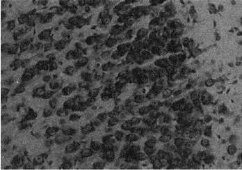
Figure 1.2 Stained cell bodies of neurons in the brain
Brain scans
Rationale
Brain imaging allows us to create pictures of a slice through the whole, living brain. Some types of scan allow for 3D images of the brain to be built up. Others allow us to get a picture of active and passive brain regions during the performance of a task.
Description
There are basically three different types of brain scan. Computerised axial tomography (CAT or CT) scans pass X-rays through the head and record sections of the brain. A series of sections can be taken to give a 3D representation of the brain. Magnetic resonance imaging (MRI) scans are similar to CT scans but instead of using X-rays they use magnetic fields. This gives a slightly clearer picture. Finally, positron emission tomography (PET) scans let us see the activity of the brain. The patient is first given an injection of a mildly radioactive form of glucose and is then put in the scanner. As those regions of the brain that are active will be using more sugar for energy, these regions will use the radioactive sugar. This radioactivity is detected by the scanner and so the active regions show up on the picture. These scanning techniques represent the major recent advance in neuroscientific investigation. They have been used, for example, to detect brain damage in certain areas of the brains of Alzheimer’s patients (Jobst 1992).
Evaluation
This method has been a major breakthrough for researchers trying to understand the workings of the normal brain and for clinicians trying to detect brain abnormalities such as tumours. The scans can also help the brain surgeon to decide whether or not surgery is necessary. The main advantage is that this is the only set of neuroanatomical methods that allow analysis in a living organism. One small disadvantage is that the machinery used to take the scans is very imposing and a few patients find the experience too traumatic (a feeling of claustrophobia is quite common). In addition, as with other methods, brain scans are very expensive to carry out.
Progress exercise Which of the techniques described so far would best be suited to investigating the following scenarios. For each scenario state the best technique and say why it would be better than the others.
- You wish to investigate where the fibres (axons) of the hippocampus project to.
- A patient presents with a suspected brain tumour.
- You are investigating the structure of the terminal bouton.
- A patient complains of reading difficulties and you wish to Investigate whether a language centre of the brain is dysfunctional (i.e. not working properly).
Neurophysiological techniques
There are several methods that allow neuroscientists to investigate how the brain works. The neuroscientist can do one of four things to investigate function. S/he can destroy part of the brain to see how the organism manages without that part of the brain (lesioning), s/he can stimulate a part of the brain to see what behaviour occurs, s/he can record from the brain to measure the activity taking place, or s/he can inject chemicals into the brain and look at the changes in behaviour that occur.
Lesioning
Rationale
By removing a part of the brain the neuroscientist can investigate that part’s function. This method can be used to test whether an area performs a particular function. It can also be used to see if an area is necessary for a function to develop. The area removed can be as small as a few millimetres in diameter or can be as large as a whole structure.
Description
Lesions are made under anaesthesia by inserting an electrode into the brain and passing a small electric current through it. The tissue around the electrode is destroyed either by the electric current itself or by the heat produced at the electrode. The size of the current determines the size of the lesion.
One example of a lesion study is by Jouvet and Renault (1966). They lesioned, in cats, an area of the brain called the raphe system and found that the cat would remain awake for a day or more. This led them to suspect that the raphe system might have a role to play in initiating sleep.
Evaluation
Lesions are very useful in helping to determine the functions of brain areas. However, care needs to be taken when interpreting the results from lesion studies. Any functional deficit may not be due to the region removed. Instead it might be because the lesion has disrupted the supply of information to that area or the supply of information from that area to another area. Another caution comes when no deficit is found after a lesion. Another, undamaged area may also (be able to) carry out the function normally reserved for the lesioned area.
Brain stimulation
Rationale
By stimulating a region of the brain it is possible to try to initiate certain behaviours and thereby learn what function(s) that region of the brain controls.
Description
Stimulation electrodes can be lowered through a small hole made in the skull. The small amount of electricity passed through is insufficient to do any damage but will stimulate the neurons in the region to fire. An example of where this has been used is to investigate pleasure centres in the brain. If a stimulating electrode is placed in an area of a...
Table of contents
- Cover Page
- Title Page
- Copyright Page
- Illustrations
- Acknowledgements
- 1: Methods of Investigating Brain Function
- 2: The Neuron
- 3: Organisation of the Central Nervous System
- 4: The Autonomic Nervous System
- 5: The Endocrine System
- 6: Homeostasis
- 7: How Drugs Affect the Brain
- 8: Classifying Psychoactive Drugs
- 9: Drugs and Behaviour
- 10: Study Aids
- Glossary
- Bibliography