This is a test
- 360 pages
- English
- ePUB (mobile friendly)
- Available on iOS & Android
eBook - ePub
The Biological Basis of Nursing: Cancer
Book details
Book preview
Table of contents
Citations
About This Book
Presenting specialized biological information on what cancer is, this book looks at how it damages the body, and how cancer treatments work. Engaging, accessible and illustrated throughout, this unique text:
- explains the basic biology of cancer
- discusses the biology of the wide range of common cancers
- identifies and explains the biological causes of cancer
- explains drug action in chemotherapy and analgesia
- explains the link between diet and cancer, and how diet is important in cancer therapy
- discusses the biological basis of a range of nursing skills linked to cancer.
Providing nurses with the essential knowledge required for working with cancer patients and their families, this book will enable them to work with current and new forms of cancer treatment.
Frequently asked questions
At the moment all of our mobile-responsive ePub books are available to download via the app. Most of our PDFs are also available to download and we're working on making the final remaining ones downloadable now. Learn more here.
Both plans give you full access to the library and all of Perlego’s features. The only differences are the price and subscription period: With the annual plan you’ll save around 30% compared to 12 months on the monthly plan.
We are an online textbook subscription service, where you can get access to an entire online library for less than the price of a single book per month. With over 1 million books across 1000+ topics, we’ve got you covered! Learn more here.
Look out for the read-aloud symbol on your next book to see if you can listen to it. The read-aloud tool reads text aloud for you, highlighting the text as it is being read. You can pause it, speed it up and slow it down. Learn more here.
Yes, you can access The Biological Basis of Nursing: Cancer by William T. Blows in PDF and/or ePUB format, as well as other popular books in Medicine & Nursing. We have over one million books available in our catalogue for you to explore.
Chapter 1
Cell biology and
cancer
- Introduction
- The cell
- The cell cycle and growth
- Tissues of the body
- The malignant cell
- A classification of malignancy
- Key points
Introduction
We are made from cells, and cells are the functional unit of life. This means that life exists at cellular level. In single cell animals (like amoeba) the one cell does everything the animal needs to live. In the human (and other multicellular animals) however, individual cells provide only a contribution towards the life of the whole being. Cells are specialised in their function, and as such each individual cell plays just a small part in the total existence of its owner. No single cell can exist without the contribution of all the others. This is both good and bad for humans. Good because a single cell animal is unable to achieve what multicellular animals can. This is because it is not possible to pack the sophisticated biochemistry that makes up a multicellular animal’s complex life into just one cell. The burden must be shared over millions of cells. By doing just that humans can build fast cars, understand the atom and walk on the moon. How many amoebae can claim those achievements? But it is also bad because failure of just a few specialised cells can jeopardise the very existence of all the others, and this is what happens in cancer.
The cell
The cell is the functional unit of the body. In humans and animals, the type of cell is known as a eukaryotic cell, whilst bacteria are prokaryotic cells (Figure 1.1). Eukaryotic cells are larger than prokaryotic cells; they have more membranes (including organelles surrounded by membranes) than prokaryotes (which do not have organelles). Bacteria have complex cell walls which eukaryotic cells do not have, and, unlike bacteria, eukaryotes have a fully formed nucleus and chromosomes.
Being multicellular, humans are made from 1014 cells (that is 100,000,000,000,000 cells). All these are derived from a single fertilised ovum. From this ovum the earliest cell divisions create a collection of cells that are all the same, but later growth in cell numbers allows the start of another process called differentiation. This means that cells begin to take on specific specialised roles, so that they will start to become tissues like blood, bone and brain. Every cell that has a nucleus also has the entire set of genes needed to make the whole body, but of course not all cells need this. The majority of genes will be switched off in most if not all cells, and only those genes needed for the cell’s specialist function will remain active.
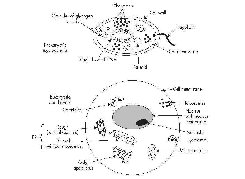
FIGURE 1.1 The prokaryote and eukaryote cells. The prokaryote cell is usually smaller with no definite nucleus, i.e. no nuclear membrane and no nucleolus. There is a cell wall in prokaryotes that eukaryotes do not have. The DNA of prokaryotes comes in a single loop, unlike the complex chromosomes found in eukaryotes. Some prokaryotes have a small additional loop of DNA called a plasmid, and some have a tail-like flagellum, both structures not found in eukaryotes. Mitochondria, endoplasmic reticulum (ER) and Golgi apparatus are features of eukaryotes.
Cells are made from protoplasm, which is mostly water with proteins, amino acids (broken down components of proteins), minerals (e.g. potassium) and other nutrients (e.g. glucose). Surrounding this protoplasm is a cell membrane (or plasma membrane) which is semi-permeable, i.e. it allows passage of some things through, including nutrients (entering the cell) and wastes (leaving the cell), and the gases oxygen (O2)(entering the cell) and carbon dioxide (CO2)(leaving the cell) (Figure 1.2). Most cells also have a nucleus surrounded by a membrane (the nuclear membrane) which is also semi-permeable, with holes of a size that allows only the passage of molecules small enough to enter or leave the nucleus, e.g. messenger ribonucleic acid (mRNA)(see p.).
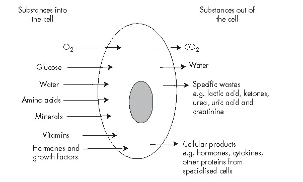
FIGURE 1.2 The input and output of the cell.
Cells have a series of organelles which carry out the various processes needed for life. In this regard, organelles are like ‘mini-organs’ (hence the term organelle), but there the comparison ends. Organs, like the heart, are made from millions of cells, but organelles are not themselves made of cells. It is not possible to have structures made from many cells inside a single cell. That would be like saying a single house brick is made from many house bricks; it just makes no sense at all. So a definition of an organelle is ‘a membrane-bound protein structure which carries out a specific function inside a cell’. Most of the organelles exist within the cytoplasm of the cell, i.e. that component of the protoplasm outside the nucleus, between the cell and nuclear membranes. One important organelle occurs inside the nucleus, the nucleolus. Table 1.1 is a list of the organelles found in a cell and their functions.
The nucleus, chromosomes and genes
The nucleus is that part of the cell that has a key central control over all cellular activities. This is because the nucleus houses the genes that code for the proteins involved is nearly all cellular functions. Genes are stretches of the molecule deoxyribonucleic acid (DNA)(Figure 1.3). Genes determine two things with regards to protein production: (1) they determine which amino acids will be in the finished protein, and (2) in what sequence these amino acids will occur. In the nucleus of human cells are found the 46 chromosomes which make up the karyotype (Figure 1.4). A chromosome is one complete molecule of DNA, and the larger chromosomes can contain many genes. The karyotype is an artificial assemblage of the chromosomes where the homologous chromosomes have been placed together in a numerical sequence from 1 to 22 (called autosomes) plus one pair of sex chromosomes. Homologous means ‘the same’ because we inherit one set of 23 chromosomes from one parent, and one set of 23 chromosomes from the other parent. So number one chromosome from the father is ‘the same’ as number one chromosome from the mother, and so on with the other chromosomes. Both number one chromosomes carry the same genes as each other, and therefore they are homologous. This means that the cell has two versions of every gene, the version from one parent and the version from the other parent. These alternative versions of the same gene are called alleles. Chromosomes certainly do not exist laid out like a karyotype inside the nucleus; they occur mixed up as separate chromatin threads. The karyotype is, however, a convenient way for us to organise chromosomes for the purpose of examination. Chromatin, the substance of chromosomes, is made from DNA and its packaging proteins called histones. Long chromatin threads are coiled to form chromatid strands. Chromatid strands are copied in the cell cycle in the S stage (see p.), and from here to anaphase of mitosis (see p.) chromosomes consist of two identical chromatid strands (called sister chromatids) joined at one point called a centromere. Deoxyribonucleic acid (DNA) has four bases mounted on a sugar-phosphate backbone (the sugar is deoxyribose). Two strands of DNA twist round each other in a double helix form and the bases are linked across from one strand to the other (see Figure 1.3). The four bases are adenine, thymine, cytosine and guanine, where adenine and thymine link together, and cytosine and guanine link together across the strands. The linkage is in the form of hydrogen bonds, two such bonds between the adenine and the thymine, three such bonds between the cytosine and the guanine. Hydrogen bonds are weak links that can be separated when the two strands come apart, and reformed when the molecule joins up again. This happens for protein synthesis and during replication of DNA (see the S phase of the cell cycle, p.).
TABLE 1.1 The cellular organelles
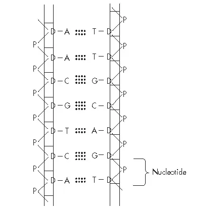
FIGURE 1.3 The DNA molecule. The sides of the molecule are made of nucleotides. Each nucleotide has a sugar (D=deoxyribose) and a phosphate (P) component with a base attached. There are four possible bases; adenine (A) links with thymine (T), and guanine (G) links with cytosine (C). Between adenine and thymine are two hydrogen bonds (two dotted lines), and between cytosine and guanine are three hydrogen bonds (three dotted lines). Many connected nucleotides create a molecule of incredible length that is coiled into a double helix.
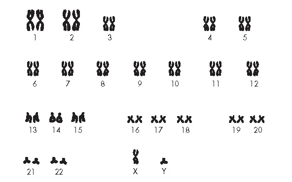
FIGURE 1.4 The human karyotype. This is an artificial presentation of the 46 chromosomes in the nucleus of the cell. Matching pairs are numbered 1 to 22 (the autosomes) and X+Y (the sex chromosomes). This is a male karyotype because it contains both X and Y. The female karyotype would contain X and X. Normally, in the nucleus, the chromosomes are jumbled and not visible, except during mitosis. (From Blows 2003, Figure 5.1.)
The cell cycle and growth
Cells grow in two ways. They grow in size (known as auxetic growth) and they grow in numbers (known as multiplicative growth). Size growth is limited because the volume of a cell (the cytoplasm that contains the metabolism) grows faster than the surface area (the cell membrane through which the nutrients and oxygen must pass) (see cell membrane, p.). Another way of putting this is to say that the volume grows by the radius cubed (radius3) whilst the surface area grows by only the square of the radius (radius2). Therefore metabolis...
Table of contents
- Cover Page
- Title Page
- Copyright Page
- Figures
- Tables
- Preface
- Chapter 1: Cell Biology and Cancer
- Chapter 2: The Causes of Cancer
- Chapter 3: Body Defences Against Cancer
- Chapter 4: Blood and Lymphatic Cancers
- Chapter 5: Cancers of the Digestive Tract
- Chapter 6: Cancers of the Respiratory and Renal Systems
- Chapter 7: Cancers In Women
- Chapter 8: Male Cancers
- Chapter 9: Metastases
- Chapter 10: Pain and Analgesia
- Chapter 11: Nutrition and the Cancer Patient
- Chapter 12: The Treatment of Cancer