eBook - ePub
General Radiography
Principles and Practices
- 258 pages
- English
- ePUB (mobile friendly)
- Available on iOS & Android
eBook - ePub
General Radiography
Principles and Practices
About this book
With chapters from globally recognized academics, General Radiography shows the multifaceted approach to general radiography and how it enhances healthcare delivery. Potentially influential to how healthcare delivery is offered, it begins with the pertinent chapters examining image acquisition and dose optimization in diagnostic radiography. Next, chapters reflect and critically discuss aspects central to patient care, and imaging within trauma, critical care and pediatric situations. The final section of this book then explores the learning, teaching and education in the field of diagnostic radiography, with novel strategies illustrated.
Tools to learn more effectively

Saving Books

Keyword Search

Annotating Text

Listen to it instead
Information
Section 1
Introductory Perspective
1 General Radiography in the Contemporary Setting
Setting the Scene
The practice of general radiography requires sound knowledge and understanding of radiographic principles, coincided with good interpersonal collaboration and person-centeredness. Looking back, I often reflect on my first experiences within this imaging modality as a radiography student, recollecting how central this imaging modality was at treating and managing a wide range of suspected health conditions within the X-ray environment. Further, I look back on the transition from film-screen (FS) to computed radiography (CR), and then digital radiography (DR). This technological change led to my own PhD study, later uncovering the effects of advancing technology and how it affected practitioners and patients alike.
General radiography, then (as the title of this book suggests) requires a more ‘generalist’ set of skills in order to undertake a wide range of projection examinations. It also hints at the possibility of general imaging being regarded as ‘non-specialist’ by convention, and thus arguably inferior when compared to other ‘specialist imaging roles’. This argument is supported by reflecting on existing job opportunities worldwide, whereby general radiographer positions are often associated with lower salaries, positioned in lower pay bandings when compared to other imaging roles affiliated with computed tomography (CT), magnetic resonance imaging (MRI), mammography and interventional imaging, for instance. My reflections of academia are also similar. Senior lecturer positions tend to seek successful applicants with ‘specialist experience’ in order to meet the demands of the ever-evolving needs of higher education institutions. This book is in part a celebration and a reminder of the virtues required to perform general radiographic examinations optimally, which remain underscored by principles and practices in both clinical and academic contexts. Henceforth, up and coming chapters reaffirm the utility of general radiographic principles and practices, which is anticipated to add value to readers transnationally.
The book begins by outlining pertinent theory regarding image acquisition and dose optimization (Section 2). The book then draws from experienced practitioners by recognizing the value of interprofessional working among radiographers within the hospital environment, and more importantly, reminds us of sustaining person-centeredness in trauma and critical care scenarios (Section 3). Finally, the editors have selected chapters concerning learning, teaching, and education in diagnostic radiography, and how virtual reality and other innovative forms of pedagogical and andragogical methods are being considered in order to enhance the delivery and experiences of undergraduate radiography students education (Section 4).
It is anticipated this book will be of use to a number of audiences. First, for student radiographers, sections 2 and 3 not only offer foundational principles but also provide experiences of delivering patient care and sound interprofessional working. Second, academics will find the content useful for knowledge transfer, while also utilizing topics for discussion. Finally, it is anticipated that postgraduate students and early career researchers will find the content and references helpful in order to define their own research objectives, and importantly uncover their own areas of original research.
In addition to the aforementioned, this book reaffirms the value of general radiography as it continues to remain the most frequent imaging modality performed worldwide, thus a useful criterion for assessing our professionalism and competency. This also raises two additional areas for discussion. First, because general radiography continues to constitute a large frequency of medical imaging examinations worldwide, it is argued that it remains important for the radiographic community to continuously challenge and reflect upon its clinical application(s). Second, while it is proffered that general radiography is perhaps seen inferior or ‘easier’ than say a ‘specialist modality’, this does not negate our need for practicing it optimally. From the author’s own observation(s), he was exposed to experimental research and sound pedagogy seeking to minimize ionizing radiation for general imaging examinations as an undergraduate student, yet an immediate disconnect was observed between classroom discussions and what was practiced clinically. For example, the use of appropriate exposure factors (Hayre, 2016) selection through phantom work, and/or the application of lead (Pb) rubber are two examples of such dichotomous practices (Hayre et al., 2017). Further, questions in the author’s own work have raised questions over person-centeredness (Hayre, Blackman and Eyden, 2016) and/or whether radiographers in the past have had the appropriate knowledge and understanding to acquire digital radiographs optimally (Hayre et al., 2017). These questions, and then subsequent findings, were not to pick holes, but to question, challenge, and remind us of our need to ensure sound radiographic competencies within an imaging modality that continues to represent the largest portion of what we do as diagnostic radiographers. In short, and regardless of technological advances an array of developmental opportunities exists as diagnostic radiographers.
General Radiography: A Platform for Innovation and Change?
The general radiography environment has historically offered a platform for both innovation and change. It is generally accepted that to practice general radiography ‘well’ requires a competent practitioner who can produce diagnostically acceptable images while ensuring that patients are treated and cared for in an altruistic manner. Further, with increasing time pressures, radiographers also require the physical skill to manoeuver X-ray equipment and obtain radiographs in an effective, but timely manner for patients and referring clinicians. While these attributes alone often ensure good radiographic outcomes, it is argued here that both innovation and organizational change play a significant role in the general imaging environment. For example, general radiography first offered abnormality detection opportunities for radiographers, commonly referred to as the ‘red dot’ system. This enabled radiographers to assist physicians by highlighting pathology (typically fractures) in the emergency department. In recent years, this has now evolved and led to the development of radiographer reporting offering a medico-legal diagnosis, a role commonly undertaken by radiologists. While some contention around the role development of radiographers exists in the literature (RANZCR, 2018, RCR, 2018, Hayre and Atutornu, 2019), the practice of radiographer reporting has become a recognized form of advanced practice, which has transcended into other imaging modalities and now become an organizational norm in most departments within the United Kingdom.
The example above is a reminder of the role general imaging plays in delivering both innovation and change, due to the emergence of new roles in order to enhance outcomes for patients. Another recent development in radiography has been that of advancing technology and the suggestion of a digital radiography champion (DRC) (Hayre et al., 2017). While this role is merely theoretical, it does aim to bridge both opportunities and challenges commonly associated with digital radiography, such as the potential for lowering ionizing radiation for patients, but also recognizing and educating radiographers of overexposure (ibid). As technology will continue to remain a significant driver for the radiography profession in years to come we may need to think outside the box in terms of delivering alternate forms of advanced practice in order to not only meet the operational needs of the department, but more importantly enhance the care and experiences of patients.
The rationale for a DRC in contemporary practice is grounded on the view that digital radiography offers significant dose optimization, without compromising image quality. However, there has also been a suggestion whether this evidence base is actually being transferred into the clinical environment and/or whether we are simply experiencing a ‘drift’ away from research evidence (Snaith, 2016), as radiographers may not always conform to the delivery of sound ionizing radiation to patients (Hayre, 2016). This offers an opportunity to bridge a theory-practice gap by means of introducing a DRC practitioner. For example, as a specialist in image acquisition and dose optimization a DRC could help connect theory and practice by undertaking and reflecting on dose audits/empirical research, deliver training and learning to staff, and conduct clinical trials in order to help enhance imaging protocols and policies with key stakeholders. Further, a DRC could become a conduit between medical physicists, referrers, radiographers, and patients by ensuring clinical questions remained radiographically sound, but also ensure that radiation doses are continuously optimized in light of technological advances.
In the past, general radiography has delivered innovative practices that have become commonplace. It is through innovation that novel opportunities can emerge, which can later lead to change and become cultural normality. For example, in the example of the DRC, this can ensure that technological advancements are optimized for patients and may allow us to rethink how ‘advanced practiced’ is considered. At present, increasing emphasis on ‘advanced practiced’ is linked to image interpretation, whereby we mimic roles performed by medical doctors. However, there is an argument that as technology continues to offer enhanced hardware and software capabilities one ongoing challenge for radiographic practitioners will be the accountability of optimizing image quality for patients, while using as little radiation as possible.
Summary
This introductory chapter offers a perspective that considers general radiography as a specialism. Although general radiography could be associated as an elementary step into the radiographic profession for radiographers, it is considered here as a modality that can offer innovation and change. First, the volume of examinations undertaken, supported with the multifaceted approach in which general radiography supports the management of patients with ill-health reflects this. There remains a wealth of opportunism for research, innovation, and change in an imaging modality that has historically demonstrated such virtues. The forthcoming chapters celebrate and reaffirm this by offering theoretical, clinical, and academic discussions with an overall aim of advancing knowledge and generating further discussion within an array of academic and clinical contexts.
REFERENCES
Hayre, C.M. (2016) ‘Cranking up’, ‘whacking up’ and ‘bumping up’: X-ray exposures in contemporary radiographic practice. Radiography, 22 (2), pp. 194–198.
Hayre, C.M. and Atutornu, J. (2019) Is image interpretation a sustainable form of advanced practice in medical imaging? Journal of Medical Imaging and Radiation Sciences, 50 (2), pp. 345–347.
Hayre, C.M., Blackman, S., Carlton, K. and Eyden, A. (2017) Attitudes and perceptions of radiographers applying lead (Pb) protection in general radiography. Radiography, 24 (1), pp. e13–e18.
Hayre, C.M. Blackman, S. Eyden, A. (2016) Do general radiographic examinations resemble a person-centred environment? Radiography, 22 (4), e245–251.
Royal College of Radiologists. The Radiology Crisis in Scotland: Sustainable Solutions Are Needed Now. [Online] Available at: www.rcr.ac.uk/posts/radiology-crisis-scotland-sustainable-solutions-are-needed-now (Accessed: 07/08/2018).
Snaith, B. (2016) Evidence based radiography: Is it happening or are we experiencing practice creep and practice drift? Radiography, 22 (4), pp. 267–268.
The Royal Australian and New Zealand College of Radiologists. Image Interpretation by Radiographers – Not the Right Solution. [Online] Available at: https://www.sor.org/system/files/news_story/201205/IPAT%20Final%20Report%2012%2034%204%2028%20(2).pdf (Accessed: 7/4/2020).
Section 2
Image Acquisition and Dose Optimization
2 Digital Receptors
Digital Image Acquisition
The launch of the now defunct National Programme for Information Technology (NPfIT) in 2002 highlighted that considerable change was required with regard to diagnostic imaging. Traditionally, images were acquired using radiographic film and the NPfIT necessitated moving from analogue to digital acquisition. This introduced the concept of Digital Radiography (DR) and technology that was able to perform the functions of radiographic film. The aim of these technologies is to convert the radiation intensities leaving the patient into electrical charge and then subsequently a binary number via an analogue to digital converter (ADC) for display and archival purposes.
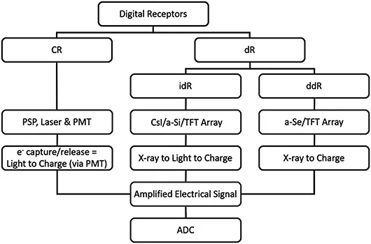
To produce images in a digital format, it was necessary to discard the use of film/screen radiography (FSR) and introduce electronically readable devices that have the ability to record the latent image. In practice, these devices are known as Digital Receptors (DRs) and take the form of Computed Radiography (CR), which uses Photostimulable Phosphor Plates (PSPs) and digital Radiography (dR), which uses Flat Panel Detectors (FPDs). There are two types of FPDs available: direct digital Radiography (ddR) and indirect digital Radiography (idR). It is important to convey here that the abbreviation DR incorporates all devices that are able to record the latent image (Figure 2.1), whereas the abbreviation dR refers to the use of FPDs to acquire an image.

Figure 2.1 Schematic Overview of Digital Receptors.
The devices used to produce digital images can be crudely thought of as a receptor that is in the form of a matrix of many individual pixels. Upon exposure to radiation intensities leaving the patient, the pixels absorb X-ray photons and the acquisition device converts the X-ray photons into an electrical signal. It can be speculated that if the X-ray photons have interacted with an area that is radiolucent, then the electrical signal is significantly large. Conversely, if the X-ray photons have interacted with an area that is radiopaque, then the electrical signal is significantly small. The electrical signal that is generated is then subsequently converted to a binary number via a...
Table of contents
- Cover
- Half Title
- Series Page
- Title Page
- Copyright Page
- Dedication
- Table of Contents
- Acknowledgments
- Preface
- Contributors
- SECTION 1: Introductory Perspective
- SECTION 2: Image Acquisition and Dose Optimization
- SECTION 3: Patient Care and Considerations
- SECTION 4: Learning, Teaching, and Education
- Index
Frequently asked questions
Yes, you can cancel anytime from the Subscription tab in your account settings on the Perlego website. Your subscription will stay active until the end of your current billing period. Learn how to cancel your subscription
No, books cannot be downloaded as external files, such as PDFs, for use outside of Perlego. However, you can download books within the Perlego app for offline reading on mobile or tablet. Learn how to download books offline
Perlego offers two plans: Essential and Complete
- Essential is ideal for learners and professionals who enjoy exploring a wide range of subjects. Access the Essential Library with 800,000+ trusted titles and best-sellers across business, personal growth, and the humanities. Includes unlimited reading time and Standard Read Aloud voice.
- Complete: Perfect for advanced learners and researchers needing full, unrestricted access. Unlock 1.4M+ books across hundreds of subjects, including academic and specialized titles. The Complete Plan also includes advanced features like Premium Read Aloud and Research Assistant.
We are an online textbook subscription service, where you can get access to an entire online library for less than the price of a single book per month. With over 1 million books across 990+ topics, we’ve got you covered! Learn about our mission
Look out for the read-aloud symbol on your next book to see if you can listen to it. The read-aloud tool reads text aloud for you, highlighting the text as it is being read. You can pause it, speed it up and slow it down. Learn more about Read Aloud
Yes! You can use the Perlego app on both iOS and Android devices to read anytime, anywhere — even offline. Perfect for commutes or when you’re on the go.
Please note we cannot support devices running on iOS 13 and Android 7 or earlier. Learn more about using the app
Please note we cannot support devices running on iOS 13 and Android 7 or earlier. Learn more about using the app
Yes, you can access General Radiography by Christopher M. Hayre, William A.S. Cox, Christopher M. Hayre,William A.S. Cox in PDF and/or ePUB format, as well as other popular books in Medicine & Medical Technology & Supplies. We have over one million books available in our catalogue for you to explore.