- 224 pages
- English
- ePUB (mobile friendly)
- Available on iOS & Android
eBook - ePub
Ultrasound and Infertility
About this book
A comprehensive survey of the use of ultrasound in management of infertile patients is presented in this publication. Particular atten-tion is given to recently developed techniques such as assessment of endometrial changes, ovarian blood flow measurements, and per-cutaneous oocyte retrieval for in vitro fertilization. The very re-cent technique of transvaginal sonography is presented and richly illus-trated with original results obtained in biopsy-guided oocyte re-trieval, and in the precise delineation of follicle size and number for infertility treatment. Guidance in the interpretation of ultrasonic findings, which include potential limitations and pitfalls, is provided in each chapter. Researchers and practitioners interested in the management of infertile patients will find this volume indispensable.
Trusted by 375,005 students
Access to over 1.5 million titles for a fair monthly price.
Study more efficiently using our study tools.
Information
Topic
MedicineChapter 1
BASIC PHYSICS OF ULTRASOUND
Branko Breyer
TABLE OF CONTENTS
I. Introduction
II. Physical Principles
A. Ultrasound Waves
B. Ultrasound Propagation in Tissues
1. Refraction and Reflection of Ultrasound Waves
III. Echoscopic Systems
A. Main Blocks of an Echoscope
B. Transducer and the Ultrasound Beam
C. Echoscope Probes and Scanning Systems
D. Attenuation Compensation: TGC
E. Dynamic Range
F. Some Notes on Resolution and Practical Use
G. Looking at the Image, Artifacts
IV. Doppler Effect and Its Use
V. Some Practical Advice
References
I. INTRODUCTION
In this chapter, we describe the basic physical and technological principles of ultrasound diagnostics without mathematical treatment, except for some simple formulas, yet include comments relevant for practical use. Ultrasound diagnostic instruments and procedures are still in fast development, so that mere knowledge of manipulation with the existing instruments is definitely insufficient for sound usage of the existing instruments to come in a few years. The knowledge of underlying principles allows one to understand what is actually new in an instrument, and what are the supposed advantages.
II. PHYSICAL PRINCIPLES
A. Ultrasound Waves
Ultrasound is, per definition, the sound of a frequency higher than the hearing limit of the human ear, i.e., above 16 to 20 kHz. Bat’s definition of ultrasound would be different. In medical diagnostics, one normally uses ultrasound waves of frequencies between 2 and 10 MHz. Basic physical principles are equally valid for audible sound as for ultrasound, only at different scales. Ultrasound is a mechanical wave, i.e., it consists of mechanical vibrations of medium particles through which it propagates. In soft tissues, the medium particles vibrate along the direction of wave propagation creating their densifications and rarefactions in space. Such a wave is called a longitudinal wave. The particles (molecules) oscillate around their (stochastic) balance positions with no net flow of matter, however, the energy flows. At very high energy densities some net flow can be induced, but this does not apply to energies of ultrasound used in diagnostics. Other types of waves like transversal and Raileigh cannot propagate to any appreciable distance in soft tissues. Ultrasound waves are characterized by parameters like frequency, wavelength, propagation speed, intensity, and pressure. Frequency is expressed in hertz (Hz), i.e., cycles per second. The physical dimension is 1/s; 1 Hz = 1 c/s, 1 kHz = 1000 c/s, and 1 MHz = 1 million c/s. The frequency used in diagnostics largely influences their properties. Wavelength is the distance between the same phases of compression of the medium in two consecutive cycles in space and is measured in meters or its subunits like millimeters. The propagation speed depends mainly on the media (tissue) properties through which the wave propagates and is related with frequency and wavelength as follows:
where c = propagation velocity in meters per second (m/s), f = frequency in hertz (Hz), and λ = wavelength in meters (m).
Strictly speaking, sounds of different frequencies travel at different velocities (frequency velocity dispersion), but these effects are negligible at frequencies, intensities, and circumstances of medical echography. The average propagation speed in soft tissues is 1540 m/s. The speed depends on both density and elastic properties of tissues.
When traveling through tissues, ultrasound causes a variation of the total pressure, i.e., the sonic pressure oscillations are superimposed upon the static pressure (atmospheric pressure). The measure of energy density flowing through a unity area in one unit of time is called intensity and is expressed in watts per square meter or centimeter (W/cm2 or W/m2).
When speaking about ultrasound intensity in an ultrasound beam, it is not sufficient to simply state the intensity, one must as well state “where” and “when”; i.e., at what position in space and within which interval. Simply stating the intensity applies only to a plane wave in a nonattenuating medium. In echography, one uses pulsed ultrasound and focused beams in attenuating media. After we discuss characteristics of ultrasound beams, we shall turn back to definitions of ultrasound intensities in the beam.
Tissue | Speed (m/s) |
|---|---|
Muscle | 1570 |
Kidney | 1560 |
Liver | 1560 |
Brain | 1510 |
Fat | 1440 |
Note: The speed values are for orientation only. Inspection of the available literature shows fairly divergent results. There is a consensus that the average speed for soft tissues should be taken as 1540 m/s.
Likewise, when speaking about frequency, one can define a single frequency only as a continuous wave. When speaking about pulses of some frequency, we usually think of frequency obtained by counting the number of zero-level crossings during the pulse. However, a pulse by physical laws contains a mixture of frequencies which we call the frequency spectrum of the pulse, and which is broader — the shorter the pulse. This fact must be born in mind in the practical use of ultrasound echography because, as we shall show, one uses different frequencies for different purposes, so it may sometimes be useful to bear in mind that when using, e.g., a probe declared 3.5 MHz central frequency, one actually transmits and receives a spectrum of frequencies, which may range from below 1 MHz to more than 5 MHz.
B. Ultrasound Propagation in Tissues
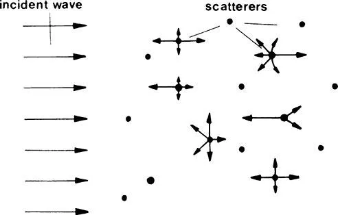
Human tissues are not homogenous in respect to ultrasound wave propagation, so that in transversing the tissues, ultrasound waves are being reflected, refracted, scattered, and absorbed. Angle of refraction depends on the respective velocities in the tissues.
The amount of reflected energy depends on characteristic acoustic impedances and angle of incidence upon the boundary of tissues. The tissues we are speaking about are different tissues in terms of elasticity and other physical properties, but can be parts of the same biological tissue. Characteristic acoustic impedance is a property of a medium and is defined as the ratio of the momentary acoustic pressure and particle velocity induced by this pressure.
In different tissues, ultrasound velocities and impedance are generally different (see Table 1). Differences between different soft tissues are much smaller than between soft tissues and bones, respectively, gas. This fact has important consequences in the use of ultrasound in medicine.
1. Refraction and Reflection of Ultrasound Waves
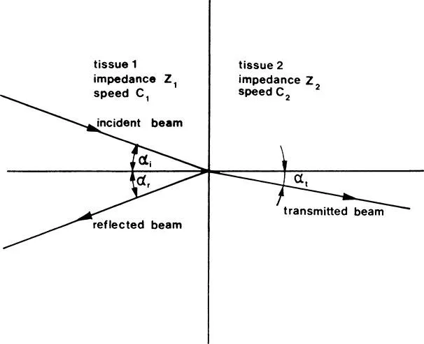
In discussing reflection and refraction of ultrasound, we shall take the simplest case — the plane wave — which, although an idealization, will give us a good idea of what happens when ultrasound waves transverse a boundary of media which is much larger than the wavelength.
As illustrated in Figure 1, a part of the energy is reflected and some of the energy is transmitted across the boundary. The angle of refraction depends on the ratio of propagation speeds in the two respective media, as described by Equation 1, while the refraction of energy reflected depends on the difference of characteristic acoustic impedances, as well as on the angle of incidence. (Equation 2a). If the speeds of ultrasound in the first and the second medium are c1 and c2, respectively, and the incidence angle is αi and the transmission angle is αt, then the refraction angle can be calculated from Equation 1:
(1) |
Equation 2 describes the ratio of the amplitudes of the reflected and the incident wave:
(2a) |
If the incidence is perpendicular to the media boundary, Equation 2a degenerates to
(2b) |
In many practical cases when we consider ultrasound waves, which are reflected back to the transmitting probe, Equation 2b gives a fairly accurate picture of what goes on.
Intensity of ultrasound is proportional to the square of its pressure so that the ratio of the reflected to the incident intensity is at 90°
(3) |
If the characteristic acoustic impedances are very different like in the case of soft tissue and gas (e.g., in lungs), from the above equation one can see that virtually all ultrasound energy will be reflected. A consequence of this is that organs containing gas, such as the lungs, cannot be examined by ultrasound, and gas in the stomach and intestines will present serious problems. Another consequence is that some sort of contact gel, oil, or other coupling agent should be applied between the ultrasound transmitter and the body to avoid air bubbles being trapped between the probe and the skin and to allow ultrasound to enter the body. It is interesting to see that ultrasound will be reflected irrespective of whether it enters from a lower or a higher impedance. The absolute value of reflected wave is the same for the same difference in characteristic acoustic impedances. On the other hand, the sign changes indicate a difference in phase.
The energy that is not reflected will be transmitted across the impedance boundary. Thus, in many cases it is the characteristic acoustic impedance that is decisive for the intensity of an echo. Sometimes the acoustic impedance is mixed up with the density by a vague analogy with X-ray imaging. This is wrong because the characteristic impedance depends on the density and propagation speed (c):
and not only on the density.
So ...
Table of contents
- Cover
- Title Page
- Copyright Page
- Preface
- Editor
- Contributors
- Table of Contents
- Chapter 1 Basic Physics of Ultrasound
- Chapter 2 Principles of Pathophysiology of Infertility Assessment and Treatment
- Chapter 3 Normal Anatomy of the Female Pelvis and Sonographic Demonstration of Pelvic Abnormalities
- Chapter 4 Ultrasonic Monitoring of Follicular Growth and Ovulation in Spontaneous and Stimulated Cycles
- Chapter 5 Transvaginal Sonography in the Management of Infertility
- Chapter 6 In Vitro Fertilization and Embryo Transfer
- Chapter 7 Interventional Ultrasound in Diagnosis and Treatment of Female Infertility
- Chapter 8 Ultrasound Doppler Studies of Blood Flow in the Pelvic Vessels
- Chapter 9 Sonography in Male Infertility
- Index
Frequently asked questions
Yes, you can cancel anytime from the Subscription tab in your account settings on the Perlego website. Your subscription will stay active until the end of your current billing period. Learn how to cancel your subscription
No, books cannot be downloaded as external files, such as PDFs, for use outside of Perlego. However, you can download books within the Perlego app for offline reading on mobile or tablet. Learn how to download books offline
Perlego offers two plans: Essential and Complete
- Essential is ideal for learners and professionals who enjoy exploring a wide range of subjects. Access the Essential Library with 800,000+ trusted titles and best-sellers across business, personal growth, and the humanities. Includes unlimited reading time and Standard Read Aloud voice.
- Complete: Perfect for advanced learners and researchers needing full, unrestricted access. Unlock 1.5M+ books across hundreds of subjects, including academic and specialized titles. The Complete Plan also includes advanced features like Premium Read Aloud and Research Assistant.
We are an online textbook subscription service, where you can get access to an entire online library for less than the price of a single book per month. With over 1.5 million books across 990+ topics, we’ve got you covered! Learn about our mission
Look out for the read-aloud symbol on your next book to see if you can listen to it. The read-aloud tool reads text aloud for you, highlighting the text as it is being read. You can pause it, speed it up and slow it down. Learn more about Read Aloud
Yes! You can use the Perlego app on both iOS and Android devices to read anytime, anywhere — even offline. Perfect for commutes or when you’re on the go.
Please note we cannot support devices running on iOS 13 and Android 7 or earlier. Learn more about using the app
Please note we cannot support devices running on iOS 13 and Android 7 or earlier. Learn more about using the app
Yes, you can access Ultrasound and Infertility by Asim Kurjak in PDF and/or ePUB format, as well as other popular books in Medicine & Gynecology, Obstetrics & Midwifery. We have over 1.5 million books available in our catalogue for you to explore.