Nuclear cardiology is critical for the medical evaluation of patients with heart disease. Clinical Nuclear Cardiology: Practical Applications and Future Directions is the second volume of this series. The volume provides information about the clinical app.
- English
- ePUB (mobile friendly)
- Available on iOS & Android
eBook - ePub
Clinical Nuclear Cardiology: Practical Applications and Future Directions
About this book
Trusted by 375,005 students
Access to over 1.5 million titles for a fair monthly price.
Study more efficiently using our study tools.
Information
Topic
MedicineSubtopic
CardiologyPositron Emission Tomography Myocardial Perfusion Imaging
Keiichiro Yoshinaga1, *, Osamu Manabe2
1 Diagnostic and Therapeutic Nuclear Medicine, National Institute of Radiological Sciences, Chiba, Japan
2 Department of Nuclear Medicine, Hokkaido University Graduate School of Medicine, Sapporo, Japan
Abstract
With the increasing availability of positron emission tomography (PET) myocardial perfusion imaging (MPI), PET MPI and the absolute quantification of myocardial blood flow (MBF) have become popular in clinical settings [1]. PET MPI shows higher diagnostic accuracy than that of single-photon emission computed tomography (SPECT) and shows predictive value for cardiac events [2, 3]. Quantitative MBF assessment also provides important additional diagnostic or prognostic information over that attained through conventional visual assessment [4]. The success of MBF quantification using PET/computed tomography (CT) has increased demand for this quantitative diagnostic approach to be more accessible. In this regard, MBF quantification approaches have been developed using several other diagnostic imaging modalities including SPECT, dynamic CT perfusion imaging, and cardiac magnetic resonance (CMR). In the United States (US), the Food and Drug Administration (FDA) has approved 13N-ammonia (13N-NH3) and 82rubidium (82Rb) for clinical use [5]. The Japanese Ministry of Health, Labour and Welfare (JMHLW) approved 13N-NH3 PET MPI for diagnosis of coronary artery disease (CAD) in March 2012 but has not approved other PET MPI tracers [6, 7]. Since 13N-NH3 PET MPI will be addressed elsewhere in this e-book, this review will address the clinical aspects of PET/CT MPI using other PET flow tracers.
Keywords: Blood Flow, Coronary Artery Disease, Endothelial Function, Flow Reserve, Positron Emission Tomography, Quantification.
* Corresponding author Keiichiro Yoshinaga: Diagnostic and Therapeutic Nuclear Medicine, National Institutes for Quantum and Radiological Science and Technology, National Institute of Radiological Sciences 4-9-1 Anagawa, Inage-Ku, Chiba, 263-8555, Japan; Tel: 81 (43) 206 3429; Fax: 81 (43) 206 4079; E-mail: [email protected]
TRACERS FOR PET MYOCARDIAL PERFUSION IMAGING
PET MPI tracers can be divided into (a) inert freely diffusible tracers such as O-15-labeled water (15O-H2O) and (b) physiologically retained tracers such as
13N-NH3, 82Rb, and 18F-flurpiridaz [8]. The basic characteristics of commonly used PET MPI tracers are listed in (Table 1).
Among these commonly used tracers, physically retained tracers can be used to produce standard tomographic images similar to those with SPECT MPI. Therefore, these tracers can be used in clinical settings. The FDA has approved these tracers for clinical use. On the other hand, it is difficult to create static images using freely diffusible tracers, and these tracers are therefore used mainly in research settings [9].
Rubidium-82
82Rb is produced from a 82strontium (82Sr) /82Rb generator which can be eluted every 10 minutes (Fig. 1) [10, 11]. Since 82Rb production does not require an on-site cyclotron, 82Rb is the most widely used tracer in PET centers [12, 13]. The physical half-life of the parent isotope is 25.5 days. Thus, the shelf life of the generator is 4 to 6 weeks. In 2016, the FDA granted additional approval of a different pharmaceutical company’s 82Sr/82Rb generator. The more recently approved generator can be used for 6 to 8 weeks and has a longer shelf life [14].
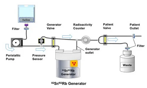
A conceptual diagram of a 82Rb infusion system that can control the 82Rb activity infusion profile. The generator valve is modulated to control the concentration of 82Rb activity administered to the patient. The radioactivity counter is used by the control system and for QA. The system can also flush the activity from the patient line at the end of the infusion. (Permission ...
Table of contents
- Welcome
- Table of Contents
- Title
- BENTHAM SCIENCE PUBLISHERS LTD.
- FOREWORD
- PREFACE
- List of Contributors
- Diagnosis of Coronary Artery Disease with Myocardial Perfusion Imaging
- Gated Myocardial Perfusion Imaging and Risk-Stratification in Coronary Artery Disease and Heart Failure
- Myocardial Viability Assessment in Predicting Prognosis and Providing Optimal Management for Ischemic Left Ventricular Dysfunction
- Cardiac Hybrid or Fusion Imaging and Future Prospective
- Positron Emission Tomography Myocardial Perfusion Imaging
- Myocardial Perfusion Imaging and other Modalities
- Fatty Acid Imaging from Basic to Clinical
- Recent Advances in BMIPP Imaging
- Potential Uses of 123mIBG and Analogous PET Tracers to Guide Use of Cardiac Implantable Electronic Devices in Heart Failure and Associated Arrhythmias
- Neurotransmitter Imaging for Cardiomyopathy and Takotsubo Syndrome
- Evaluation of Cardiac Sympathetic Nerve Function using Iodine-123 Metaiodobenzylguanidine Scintigraphy
- Standardization of 123I-meta-iodobenzylguanidine Heart-To-Mediastinum Ratio: Technical Aspects
- Image Diagnosis of Arteriosclerosis and Possibility of Early Intervention
- Nuclear Cardiology for the Diagnosis of Takotsubo Syndrome
- Small Rodent Animal Positron Emission Tomography/Single Photon Emission Tomography
Frequently asked questions
Yes, you can cancel anytime from the Subscription tab in your account settings on the Perlego website. Your subscription will stay active until the end of your current billing period. Learn how to cancel your subscription
No, books cannot be downloaded as external files, such as PDFs, for use outside of Perlego. However, you can download books within the Perlego app for offline reading on mobile or tablet. Learn how to download books offline
Perlego offers two plans: Essential and Complete
- Essential is ideal for learners and professionals who enjoy exploring a wide range of subjects. Access the Essential Library with 800,000+ trusted titles and best-sellers across business, personal growth, and the humanities. Includes unlimited reading time and Standard Read Aloud voice.
- Complete: Perfect for advanced learners and researchers needing full, unrestricted access. Unlock 1.5M+ books across hundreds of subjects, including academic and specialized titles. The Complete Plan also includes advanced features like Premium Read Aloud and Research Assistant.
We are an online textbook subscription service, where you can get access to an entire online library for less than the price of a single book per month. With over 1.5 million books across 990+ topics, we’ve got you covered! Learn about our mission
Look out for the read-aloud symbol on your next book to see if you can listen to it. The read-aloud tool reads text aloud for you, highlighting the text as it is being read. You can pause it, speed it up and slow it down. Learn more about Read Aloud
Yes! You can use the Perlego app on both iOS and Android devices to read anytime, anywhere — even offline. Perfect for commutes or when you’re on the go.
Please note we cannot support devices running on iOS 13 and Android 7 or earlier. Learn more about using the app
Please note we cannot support devices running on iOS 13 and Android 7 or earlier. Learn more about using the app
Yes, you can access Clinical Nuclear Cardiology: Practical Applications and Future Directions by Shinro Matsuo in PDF and/or ePUB format, as well as other popular books in Medicine & Cardiology. We have over 1.5 million books available in our catalogue for you to explore.