eBook - ePub
Chiropractic Radiography and Quality Assurance Handbook
- 512 pages
- English
- ePUB (mobile friendly)
- Available on iOS & Android
eBook - ePub
Chiropractic Radiography and Quality Assurance Handbook
About this book
Chiropractic Radiography and Quality Assurance Handbook is the first book devoted to erect and recumbent radiographic positioning and a practical approach to quality assurance and radiographic quality control testing. It provides a step-by-step approach to performing quality radiographic studies using radiographic images to demonstrate placement of anatomical markers and the safest location for patient identification information.
Some topics covered include:
o The importance of sound radiation safety practices and appropriate protection and collimation
o Spinal radiography including changes in positioning to reduce exposure to female patients
o Extremity radiography, covering common and specialty views to assist in diagnosis of sports injuries.
Designed for both the practitioner and the student, this book provides all of the tools necessary to produce quality radiographs in a quick reference, detailed, step-by-step approach to positioning. And adding information about darkroom and film storage, film processing quality control, film artifact identification and problem solving, makes this is an in-depth, authoritative guide.
Trusted by 375,005 students
Access to over 1.5 million titles for a fair monthly price.
Study more efficiently using our study tools.
Information
Topic
MedicineChapter | 1 |
Basic Radiography Concepts and Principles |
The first three sections of this chapter review some of the key properties of radiographic image production. The first section discusses the equipment and accessories needed to produce consistently high-quality radiographs. It also covers shielding and some recommendations on room design. The second section is a review of radiation physics principles that impact image quality. The third section introduces the elements of a technique chart and discusses more physics.
The remaining sections of the chapter explore the darkroom and film processing. The first time a student enters the darkroom, it can very intimidating. There are some important safety issues for working near and with processing chemicals. There are environmental issues about the handling of used processing solutions, which is also covered in this chapter. Samples of the Material Data Safety Sheets for Developer and Fixer are included. Later in the Quality Control chapter (Chapter 21), mechanical sections of an automatic film processor are covered.
1.1 Chiropractic Radiographic Equipment and Accessories
The radiographic equipment and accessories needed to properly perform chiropractic radiography should be as modern as the doctor can afford. The greatest positioning skill will not overcome the limits of old and poorly maintained equipment. The chiropractor should consider the age and body habitus of potential patients. One must also consider if one is intending to do radiographic studies other than basic limited spinal radiography. Chiropractic radiography is more difficult to do compared to generally recumbent medical radiography. The patient will be much less stable. The patient will also be farther away from the film so loss of resolution due to increased object to film distance will impact the film quality. Good erect radiography requires top-quality equipment and very precise positioning of the patient.
The X-ray Machine
One of the greatest deterrents of good image quality is patient motion. If limited to a single-phase machine, one is twice as likely to get motion on the film than when using a high-frequency or three-phase machine. This is due to the single-phase unit being less efficient and having limited selection of mA and time selection. Because the typical lateral view of the lumbar spine has an optimum kVp of 90 kVp, it is not uncommon to have exposure times well in excess of 1 second. For long exposure times, even involuntary body motion such as peristalsis can result in a repeated film.
It is difficult to keep older equipment in proper calibration. If one wants to use 30 mAs for a film, the exposure should be the same on the large or small focal spot, and from mA station to mA station. As the X-ray tube ages, tungsten is built up on the window of the tube. This adds filtration to the beam and reduces contrast on the film. The filament will be the first part of the tube to show age. This impacts the geometric detail of the film.
When first setting up an X-ray department, have a physicist or other qualified person test the equipment before paying for it. This is extremely important with used equipment or when buying a practice. Used X-ray units that are not high frequency or three phase should definitely be evaluated before purchase. It might be better to wait until the practice grows and buy new equipment rather than install someone’s obsolete equipment.
The grid in the film holder or Bucky must not be overlooked. One will need at least 103 lines and a 10:1 ratio to do adequate-quality spinal radiography. Make sure that it is installed perpendicular to the central ray. Many units in offices get grid cut-off because the grid is not properly aligned to the tube. Also make sure that the focal distance of the grid is consistent with the studies that will be done in the office.
One should have a Non-Bucky film holder for the erect film holder. The lateral cervical spine should never be taken using the grid at 72 in. SID; one is overexposing the patient by 5 times compared to the use of the Non-Bucky film holder. That overexposure also reduces the life of the X-ray tube.
There will be occasions when erect radiography is not practical, particularly with elderly or handicapped patients. A mobile X-ray table can be a very valuable part of the X-ray equipment. The alternative may be to put the patient on the floor to do recumbent studies. Examinations such as lumbar oblique view are best done recumbent. With any lordotic curve, it is impossible to get the lumbar spine in the same plane when doing obliques erect. For obese patients, the adipose tissue will roll away from the spine when X-rayed recumbent.
The basic X-ray room should include a three-phase or high-frequency generator, a Bucky with a 10:1 or better grid, and a mobile X-ray table (see Figure 1.1). The room must be shielded to 7 feet above the floor. The controls must be shielded and have a leaded glass window to observe the patient during the study. The equipment should be serviced according to manufacturer recommendations. A systematic quality assurance program should monitor the performance of the equipment.
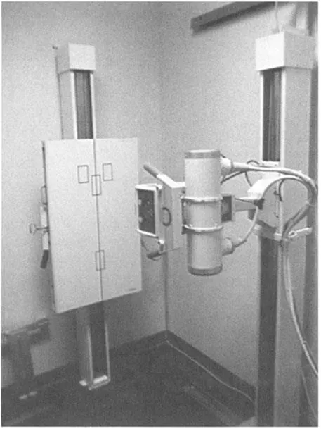
Basic X-ray unit.
The Film and Cassettes
Each component must be matched to the other components. All brands of film are not created equal. Film brands cannot be switched from one type of cassette and screen to another without the potential for grave problems. Some screens generate a blue/green spectrum of light, while others produce a green spectrum. If the film is not matched to the light spectrum of the screens, the potential for significant overexposure to the patient can result. The Relative Speed Value (RSV) is used to establish the exposure parameters of a given screen and film combination. It works very similar to the speed system used for photographic film.
The standard of the radiographic community for general radiography is a 400 speed screen and film combination. These are generally green-spectrum systems. This is a relatively high speed and low exposure system. All film manufacturers make this speed. The quantum mottle or noise on the film is acceptable and it will provide good detail for general Bucky-type work. Some manufacturers make a relatively good 800 speed system; it is noisier than the 400 speed system. There are systems as high as 1600 but the noise or grainy image makes them of limited value. A very high-speed screen and film combination is not a substitute for a more powerful X-ray machine. If planning to use a blue-sensitive system, such as “high speed” or” high speed plus” (200 speed) for general radiography, one can reduce exposure and exposure times with a 400 speed system.
Do not attempt to do small extremity work without a dedicated extremity cassette system. The 80 speed Kodak Lanex Fine system uses the same film as the 400 speed Lanex Regular system. This makes their use very practical. Some of the extremity systems use single emulsion film that increases inventory costs. The 400 speed systems are too fast for hand, ankle, foot, and phalange studies. The kVp used with 400 speed systems is usually way too low and the contrast so poor that it is difficult to avoid overexposure. With extremities, one also needs finer detail that is not available with the 400 speed system.
The Kodak Lanex Cassette System provides very good screen contact and consistent image quality using Kodak and other manufacturers’ film. They do not last forever. In 10 years of use, they will start to crack at the hinges. Proper care of the cassettes is very important to get full useful life from them. (See Figure 1.2.)
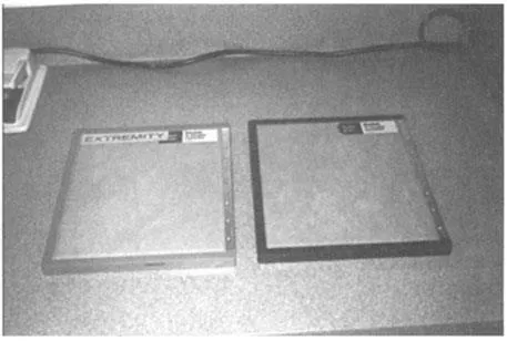
Cassette types.
Accessories and Radiation Protection
Obtain some positioning sponges and lead blockers. They will make it easier and more comfortable to position the patient. Anatomical and view lead markers must also be used. One can get by with just right and left markers and an arrow.
Gonad protection must be provided. This should include a half-apron of 0.5 mm lead rating. All aprons should be tested at least annually for holes and leaks. If one plans to take films on children or take stress films, lead gloves and a full-coat apron will be needed for one’s own protection.
Compensating Filters
Full spine radiography should not be attempted without compensating filters, such as those manufactured by Dr. Ralph Nolan. Thes...
Table of contents
- Cover
- Title Page
- Copyright Page
- Author
- Preface
- Acknowledgement
- Table of Contents
- Chapter 1 Basic Radiography Concepts and Principles
- Chapter 2 Radiation Safety in Chiropractic Radiography
- Chapter 3 Developing a System or the Proper Sequence for Taking Films
- Chapter 4 Spinal Positioning Quick Reference Charts
- Chapter 5 Cervical Spine Radiography
- Chapter 6 Thoracic Spine, Chest, and Bony Thorax Radiography
- Chapter 7 A-P Full Spine and Lumbar, Sacrum, Coccyx Radiography
- Chapter 8 Abdominal and Chest Decubitus Radiography
- Chapter 9 Basic Skull and Facial Radiography
- Chapter 10 Introduction to Extremity Radiography
- Chapter 11 Extremity Positioning Quick Reference Charts
- Chapter 12 Hand and Wrist Radiography
- Chapter 13 Forearm, Elbow, and Humerus Radiography
- Chapter 14 Shoulder, Scapula, and Clavicle Radiography
- Chapter 15 Pelvis, Hips, and Femur Radiography
- Chapter 16 Knee and Patella Radiography
- Chapter 17 Lower Leg and Ankle Radiography
- Chapter 18 Calcaneus, Foot, and Toe Radiography
- Chapter 19 Introduction to Quality Assurance and Radiographic Quality Control
- Chapter 20 Darkroom and Film Storage Quality Control
- Chapter 21 Film Processing Quality Control
- Chapter 22 Radiographic Quality Control Testing
- Chapter 23 Accessory Quality Control Testing
- Chapter 24 Film Artifact Identification and Problem-Solving
- Appendix 1 Review of Basic Radiographic and Quality Control Concepts
- Appendix 2 A Review to Assist in Interpretation of Radiography with Common Film Marking Lines
- Appendix 3 Common Radiography Terms, Rules, and Rare-Earth Screen and Film Combination Chart
- Index
Frequently asked questions
Yes, you can cancel anytime from the Subscription tab in your account settings on the Perlego website. Your subscription will stay active until the end of your current billing period. Learn how to cancel your subscription
No, books cannot be downloaded as external files, such as PDFs, for use outside of Perlego. However, you can download books within the Perlego app for offline reading on mobile or tablet. Learn how to download books offline
Perlego offers two plans: Essential and Complete
- Essential is ideal for learners and professionals who enjoy exploring a wide range of subjects. Access the Essential Library with 800,000+ trusted titles and best-sellers across business, personal growth, and the humanities. Includes unlimited reading time and Standard Read Aloud voice.
- Complete: Perfect for advanced learners and researchers needing full, unrestricted access. Unlock 1.5M+ books across hundreds of subjects, including academic and specialized titles. The Complete Plan also includes advanced features like Premium Read Aloud and Research Assistant.
We are an online textbook subscription service, where you can get access to an entire online library for less than the price of a single book per month. With over 1.5 million books across 990+ topics, we’ve got you covered! Learn about our mission
Look out for the read-aloud symbol on your next book to see if you can listen to it. The read-aloud tool reads text aloud for you, highlighting the text as it is being read. You can pause it, speed it up and slow it down. Learn more about Read Aloud
Yes! You can use the Perlego app on both iOS and Android devices to read anytime, anywhere — even offline. Perfect for commutes or when you’re on the go.
Please note we cannot support devices running on iOS 13 and Android 7 or earlier. Learn more about using the app
Please note we cannot support devices running on iOS 13 and Android 7 or earlier. Learn more about using the app
Yes, you can access Chiropractic Radiography and Quality Assurance Handbook by Russell L. Wilson,Russell Wilson in PDF and/or ePUB format, as well as other popular books in Medicine & Alternative & Complementary Medicine. We have over 1.5 million books available in our catalogue for you to explore.