eBook - ePub
Angiography and Plaque Imaging
Advanced Segmentation Techniques
- 560 pages
- English
- ePUB (mobile friendly)
- Available on iOS & Android
eBook - ePub
Angiography and Plaque Imaging
Advanced Segmentation Techniques
About this book
Recent, rapid advances in mathematical engineering and applied mathematics have opened the door to solving complex problems in angiography imaging. For the first time, this book presents the different medical imaging modalities--MR, CT, x-ray, and ultrasound--for performing angiography and its analysis. Pioneers from a variety of relevant disciplin
Tools to learn more effectively

Saving Books

Keyword Search

Annotating Text

Listen to it instead
Information
1
Non-Skeleton- and Skeleton-Based Segmentation Techniques from Angiography Data Sets
Jasjit S. Suri, Kecheng Liu, Sameer Singh, and Swamy Laxminarayan
CONTENTS
1.1 Introduction
1.2 A Brief Note on MRA Data Acquisition and Prefiltering
1.3 Non-Skeleton (Direct) Vascular Segmentation Techniques
1.4 Skeleton (Indirect) Vascular Segmentation Techniques
1.5 Discussions of Skeleton-Based vs. Non-Skeleton-Based Methods
1.6 Challenges, the Future, and Summary: Vascular Segmentation
Acknowledgments
References
1.1 Introduction
Vascular diseases are one of the major sources of deaths in the United States. A report [1] states that “Aneurysm rupture is not a rare occurrence as evidenced by the fact that this event currently ranks thirteenth on the list for leading causes of death in the U.S.A.” Also, this report [1] says that “Chronic venous insufficiency is a common problem in the U.S., affecting approximately 5% of the general population. It is estimated that half a million patients suffer from ulceration of the lower extremity as a result of longstanding venous disease.” This report raises the importance of research needed in the area of angiography, the branch of medicine that deals with veins and arteries. In the field of cerebrovascular diseases [1], “Ischemic and hemorrhagic stroke account for one of the principal causes of death and disability in older aged population. In fact, stroke currently ranks third on the list of leading causes of death in the United States (see also [2–5]).
Each year, approximately 500,000 people suffer a new or recurrent stroke in the U.S., and approximately 150,000 die as a result of this process. In addition, more than one third of stroke survivors are left with permanent physical disability, which currently accounts for the leading cause of nursing home admissions in the U.S.”
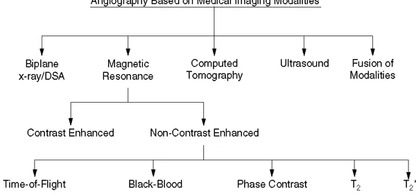
FIGURE 1.1 This chart shows the various ways angiography can be performed, based on the type of medical imaging modality used. Note that T2 and T2. stand for T2-weighted and T2.-weighted imaging.
Having discussed the national concern on vascular diseases, we briefly present different imaging modalities that can perform angiography. figure 1.1 shows the different ways angiography can be performed. The main modalities include biplane x-ray/digital subtraction angiography (DSA) (see [6–9]); MR angiography (MRA) (see [10–19]); CT angiography (CTA) (see [20–25]); ultrasound angiography1 (see [26–32]); and angiography as a fusion of different medical imaging modalities (see [33–35]). From the clinical point of view, DSA2 is considered the most reliable and accurate method for vascular imaging, a subset of x-ray. On the contrary, this method lacks three-dimensional information,3 which is easily available via MR and CT techniques. The MR and CT techniques, however, lack the ability to locate tiny vessels and the morphological estimation of stenoses and aneurysms.
With advancements in MR technology and MR acquisition techniques,4 the burden on vascular segmentation has certainly been reduced. But due to the complexity of the body vasculature, occlusions, small diameters of the vessels, and other factors, the segmentation problem has not been fully solved. Researchers are aggressively attempting to segment the vessels (arteries and veins) from the human body using computer vision, graphics, and imageprocessing (CVGIP) techniques and recently there has been tremendous interest in using automated segmentation of vasculature from MRA data sets. The main motivations for performing vascular segmentation include:
- Neuro-surgical planning, interventional procedures and treatments. The ability to perform segmentation of three-dimensional brain vessels is important for neurosurgeons because it aids in the visualization of “intracranial saccular aneurysms” in relation to the vessels (see [36–39]). This helps in planning the brain surgery and in performing angioplasty. The position of the tumor can be localized using the three-dimensional venous structures as landmarks or roadmaps before and during the surgery (see [37]). Similar examples can be seen in the abdomen, such as for abdominal aortic aneurysms (AAA). Segmentation also helps in real-time operating room decision making and post-operative monitoring. Segmentation of the vasculature can help in endovascular treatment for intracranial saccular aneurysms (see [40]).
- Time saving. Automated or semi-automated vasculature segmentation helps surgeons, radiologists, and oncologists in many different ways. For example, it can save radiologists a significant amount of time required for manual segmentation and can facilitate further data analysis. Another example is the preoperative evaluation of patients scheduled for endovascular repair by performing the computation of the minimum and maximum diameters of the lumen. These measurements are acquired in the planes perpendicular to a previously determined central lumen line at certain intervals.
- Distinguishing between the veins and arteries. Segmentation also helps the radiologists and physicians in separating and visualizing arteries and veins (bright5 blood and dark6 blood vessels) (see Figure 1.4 and Figure 1.5).
- Relative placement of the brain structures. Segmenting brain areas and brain blood vessels helps surgeons in studying the growth process of the brain structures and their relationships to each other.
- Blood flow process and hemodynamics. Research is currently active in studying the relationship of the blood flow7 in the blood vessel structures (see Figure 1.7). This includes hemodynamics and wall shear stress in the development and progression of atherosclerosis and other vascular diseases (see [41–43]). Wall shear stress measurements in the abdominal aorta and other body parts need to be further researched (see [44]).
- Stenosis and aneurysm assessment/quantification. Segmentation of vasculature is very useful for the detection and grading of stenoses and aneurysms and for assessment (see [45–49]), especially in the areas of brain, bronchial trees, and bile ducts. This is particularly helpful in the clinical evaluation of atherosclerosis. The shape, size, and location of aneurysms are critical in the embolization procedure.8
- Disease monitoring/remission. Having an accurate description of vessel structures is important for monitoring disease progress or remission.
- Quantification of vascular structures. Since the centerline vessel axis can serve as a basis for the description of quantitative diagnosis, vessel quantification is necessary (see [38]).
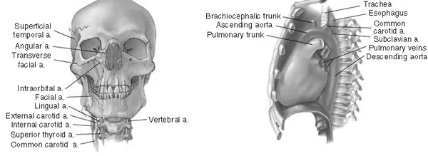
FIGURE 1.2 (See color insert.) Vasculature system for the head and neck and the heart embedded in the skull and chest cavity, respectively. Left: Neck arteries entering the skull area. Right: Aorta seen in the chest cavity. (Courtesy of Professor Fishman, Johns Hopkins University, Baltimore, MD).
Having discussed the applications and sources of motivations for performing vascular segmentation, we now present the major difficulties in performing segmentation of the vasculature in MR data sets. The following are the main reasons contributing to the complexity in vasculature segmentation from MRA data sets (see [18]):
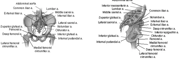
FIGURE 1.3 (See color insert.) Vasculature system for pelvis region. Left: Pelvis frontal view. Right: Pelvis lateral view. (Courtesy of Professor Fishman, Johns Hopkins University, Baltimore, MD).
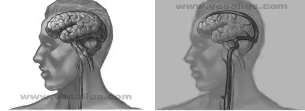
FIGURE 1.4 (See color insert.) Right: The brain and its carotid artery suppl...
Table of contents
- Cover Page
- Title Page
- Dedication
- Preface
- Acknowledgments
- The Editors
- Contributors
- 1: Non-Skeleton-and Skeleton-Based Segmentation Techniques from Angiography Data Sets
- 2: Scale-Space 3-D Ellipsoidal Filtering for White and Black Blood MRA
- 3: Three-Dimensional Interactive Vascular Postprocessing Techniques: Industrial Perspective
- 4: Masking Strategies for Black-Blood Angiography
- 5: Segmentation of Retinal Fundus Vasculature in Nonmydriatic Camera Images Using Wavelets
- 6: Automated Model-Based Segmentation, Tracing, and Analysis of Retinal Vasculature from Digital Fundus Images
- 7: Atherosclerotic Plaque Imaging Techniques in Magnetic Resonance Images
- 8: Analysis and Visualization of Atherosclerotic Plaque Composition by MRI
- 9: Plaque Morphological Quantitation
- 10: On the Role of Computer Vision in Intravascular Ultrasound Image Analysis
- 11: Clinical Applications Involving Vascular Image Segmentation and Registration
- 12: A Note on Future Research in Vascular and Plaque Segmentation
- Key Words
Frequently asked questions
Yes, you can cancel anytime from the Subscription tab in your account settings on the Perlego website. Your subscription will stay active until the end of your current billing period. Learn how to cancel your subscription
No, books cannot be downloaded as external files, such as PDFs, for use outside of Perlego. However, you can download books within the Perlego app for offline reading on mobile or tablet. Learn how to download books offline
Perlego offers two plans: Essential and Complete
- Essential is ideal for learners and professionals who enjoy exploring a wide range of subjects. Access the Essential Library with 800,000+ trusted titles and best-sellers across business, personal growth, and the humanities. Includes unlimited reading time and Standard Read Aloud voice.
- Complete: Perfect for advanced learners and researchers needing full, unrestricted access. Unlock 1.4M+ books across hundreds of subjects, including academic and specialized titles. The Complete Plan also includes advanced features like Premium Read Aloud and Research Assistant.
We are an online textbook subscription service, where you can get access to an entire online library for less than the price of a single book per month. With over 1 million books across 990+ topics, we’ve got you covered! Learn about our mission
Look out for the read-aloud symbol on your next book to see if you can listen to it. The read-aloud tool reads text aloud for you, highlighting the text as it is being read. You can pause it, speed it up and slow it down. Learn more about Read Aloud
Yes! You can use the Perlego app on both iOS and Android devices to read anytime, anywhere — even offline. Perfect for commutes or when you’re on the go.
Please note we cannot support devices running on iOS 13 and Android 7 or earlier. Learn more about using the app
Please note we cannot support devices running on iOS 13 and Android 7 or earlier. Learn more about using the app
Yes, you can access Angiography and Plaque Imaging by Jasjit S. Suri, Swamy Laxminarayan, Jasjit S. Suri,Swamy Laxminarayan in PDF and/or ePUB format, as well as other popular books in Medicina & Biotecnologia in medicina. We have over one million books available in our catalogue for you to explore.