eBook - ePub
Stem Cells
Scientific Facts and Fiction
- 324 pages
- English
- ePUB (mobile friendly)
- Available on iOS & Android
eBook - ePub
Stem Cells
Scientific Facts and Fiction
About this book
Recent advances in the fields of medicine and technology have led to the development of stem cell therapy. A stem cell is a cell that has the potential to develop into many different types of cell in the body. It has the ability to divide and copy itself and at least one other specialized type of cell.
Stem Cells was written to provide information about the development of stem cell therapy, which can be used in the fields of research and medicine. The main goal of the book is to provide readers with an overview of the scientific facts about stem cells and its promising effects on the human body, as well as on the creation of new drugs and medicines. The book also highlights the ongoing clinical research into stem cells and lists the therapies whose effectiveness is being investigated.
Many scientists argue that stem cell therapy will be of great help to patients and society if it is proven to be safe and effective.
- Explains in straightforward, non-specialist language the basic biology of stem cells and their applications in modern medicine and future therapy
- Includes extensive coverage of adult and embryonic stem cells both historically and in contemporary practice
- Richly illustrated to assist in understanding how research is done and the current hurdles to clinical practice
Trusted by 375,005 students
Access to over 1.5 million titles for a fair monthly price.
Study more efficiently using our study tools.
Information
Subtopic
Cell BiologyIndex
Biological SciencesChapter 1
The Biology of the Cell
Outline
Organisms are Composed of Cells
DNA, Genes and Chromosomes
How is the Amount of mRNA Regulated?
Transcription Factors
Epigenetic Regulation
RNA Interference
From mRNA to a Functional Protein
From DNA and Proteins to a Cell with a Function …
DNA Differences Between Genomes: Mutation or Variation?
Diseases Due to Variations and Genome Mutations
Dominant or Recessive?
DNA Outside the Nucleus: Bacterial Remains
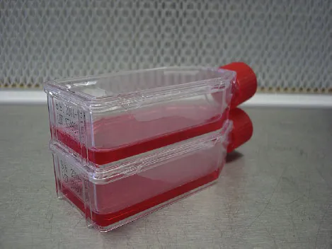
Cell Lines and Cell Culture
This book is about stem cells. Stem cells and their applications in clinical medicine, biotechnology and drug development for pharmaceutical companies, involve many facets of biology; from genetics and biochemistry to synthetic scaffolds and three-dimensional architecture for tissue engineering. For this reason the most important molecular and cell biological principles needed to understand stem cells will be introduced to the reader in this chapter.
Organisms are Composed of Cells
Humans and animals, as well as plants and trees, contain many different functional organs and tissues. These, in turn, are composed of a large variety of cells. Cells are therefore the basic building blocks which make up the organism. All animal cells have a similar structure: an outer layer called the plasma membrane, made up of a double layer of lipid molecules, and an inner fluid known as cytoplasm. The cytoplasm contains a variety of small structures called organelles, each of which has a specific and essential function within the cell. Most cell organelles are themselves separated from the cytoplasm by their own membranes. The form of the cell is determined and supported by the cytoskeleton, a flexible scaffolding composed of polymers of protein molecules which form a network that shapes the cell and allows it to move and “walk”. Inside the cell countless proteins – sometimes in cooperation with RNA molecules – facilitate the chemical and physical reactions and transport of other molecules required to carry out specific cellular functions.
The most prominent organelle viewed under the microscope is the nucleus. This contains the chromosomes, which are in part made up of DNA – one long molecule of DNA per chromosome – representing the
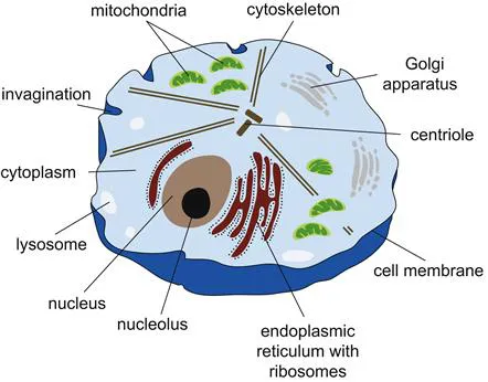
Schematic representation of an animal cell. The cell contains a fluid called the cytoplasm, enclosed by a cell membrane. The nucleus contains the genetic information, the DNA. The shape of the cells is determined by their cytoskeleton. Proteins and lipids are generated and assembled in the endoplasmic reticulum. The Golgi apparatus is then responsible for further transport within the cell. Lysosomes are small vesicles with enzymes that can break down cellular structures and proteins that are no longer required, while the energy necessary for the cell is generated by the mitochondria.
organism’s blueprint. Although cells can have different shapes and functions, the DNA sequence in all cells of a given individual is in principle identical (with the exception of certain blood cells). Other prominent structures in the cell are the mitochondria. These organelles are present in large numbers and generate the energy required by the cell. Cells with very large energy requirements, like heart cells, contain correspondingly higher numbers of mitochondria. Energy is also required, among other things, for creating messenger RNA (mRNA) and linking amino acids to each other in the ribosomes to form proteins. Rudimental proteins thus made are delivered to the tubular structures of the endoplasmic reticulum, where they are processed into actual working proteins in yet another organelle, the Golgi apparatus. They are then transported in small vesicles, termed vacuoles, to the site in the cell where they are required to exert their own specific function. Each cell is thus a highly dynamic structure with its own powerhouse, factories and transport systems.
1.1 ANTONI VAN LEEUWENHOEK
Although plants and animals have been studied for centuries, the realization that organisms are composed of cells is of relatively recent origin. This is simply due to the fact that in the past there were no technologies available for cells to be seen. It was only after the invention of the microscope (around 1595) that cells could be made visible for the first time.
The Dutchman Antoni van Leeuwenhoek was one of the first microscopists; he dedicated himself to the discovery and description of the hitherto invisible world of biology. Van Leeuwenhoek was born on October 24, 1632 in Delft, The Netherlands. Quite unlike other great scientists of his day, he did not receive a university education, but was entirely self-taught. His naïve approach, disregarding any form of scientific dogma, allowed him to think freely and be guided only by his own enthusiasm and interest.
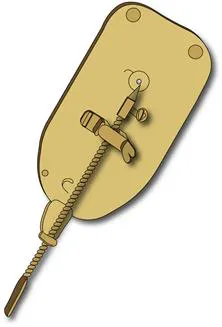
The microscopes made by van Leeuwenhoek were in fact magnifying glasses of outstanding quality.
Antoni van Leeuwenhoek was by trade a salesman in household linen and used magnifying glasses to examine the quality of cloth. He ground his own lenses using diamond shavings, which he obtained from Delftdiamond cutters. He also built his own microscopes, basically simple instruments containing a single lens, but ground with high precision, sufficient to achieve magnifications of around 300×. van Leeuwenhoek’s microscopes consisted of two metal plates fixed to each other with a lens between them. The lens was fixed and the object to be examined was placed on top of a metal holder that could be moved using a set-screw, while focusing occurred through a screw at the back of the instrument. The entire construction was less than 10 cm in size. van Leeuwenhoek’s microscopes were in essence only very strong magnifying glasses, quite different from the composite microscopes that also existed at the time. However, it was his curiosity and insight – combined with the quality of the lenses and his ability to illuminate the objects properly – that allowed him to discover the microscopic world. He examined water from ditches, tooth plaque, baker’s yeast, stone dust, blood and sperm.
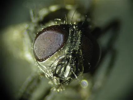
Antoni van Leeuwenhoek studied many small objects with his microscope, among which were the composite eyes of insects, and tried to determine the number of facets on each eye.
The Delft physician Reinier de Graaf introduced van Leeuwenhoek to the Royal Society in London, after which he published his findings in a total of 200 letters (in Dutch) which he sent to the Society. The letters, which needed to be translated into English or Latin for publication, are anecdotal, containing a panoply of random observations, but uniquely detailed in their descriptions. van Leeuwenhoek achieved international fame with his observations, but in a letter written in 1716 he said that he “did not strive for fame, but [was] driven by an inner craving for knowledge”. This drive he believed to be stronger in him than in most other people. van Leeuwenhoek died on August 16, 1723.
DNA, Genes and Chromosomes
The DNA (deoxyribonucleic acid) in each cell of our body contains all of the information needed to create a complete individual. In humans, DNA is divided into around 23,000 different genes, each of which encodes the blueprint for one or more proteins. What does the information in the DNA look like, and how is it translated into the production of proteins? How does the cell decide which proteins to make?
A DNA strand is composed of a long series of nucleotides. Each nucleotide consists of a deoxyribose molecule, which forms the backbone of the DNA molecule, and is linked to one of four bases: adenine (A), guanine (G), thymine (T) and cytosine (C). Nucleotides are interconnected by phosphate groups, forming a long chain. The specific sequence of the different bases represents the core of the DNA code. Two single strands of DNA combine to form a double-stranded DNA molecule as complementary bases form base-pairs held together through hydrogen bridges: adenine binds to thymine, while guanine always binds to a cytosine.
Normally, the DNA in a chromosome is double-stranded and forms a long chain. These long double-stranded DNA molecules (one DNA molecule can be up to 10 cm long) form the famous Watson and Crick DNA “double helix” structure, itself wrapped around a core of a special family of proteins called histones. The histone proteins provide the extremely long DNA molecule with the support and guidance to fold into a complicated three-dimensional form, which fits into the small space of the nucleus and enables correct use of each part of the DNA molecule. In the nucleus, this intricate combination of DNA and proteins is called chromatin. The ends of the DNA molecules which form the caps of the chromosomes are called telomeres. These telomeres protect the chromosome ends from DNA damage but are shortened after each cell division.
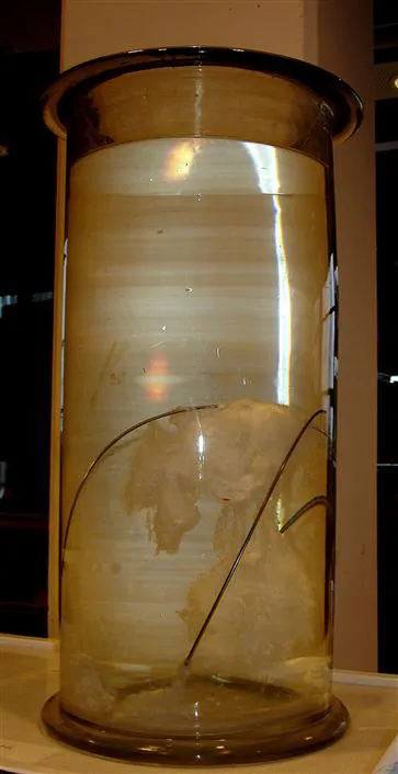
DNA can be isolated from cells and “precipitated” (separated from liquid). It then appears as a white glue-like substance. This meter-high glass pot contains the same amount of DNA as would be present in all of the cells of an adult human.
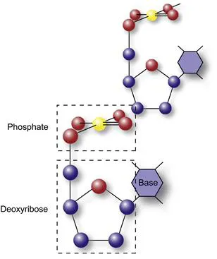
A single DNA strand is a long polymer composed of sugar (deoxyribose) and phosphate groups that together form the backbone of the DNA. Either one of the possible four bases adenine, thymine, cytosine or guanine are coupled to the deoxyribose strand.
Cells can continue dividing until the telomeres are “used up”; this is the process of cellular aging or senescence.
The number of chromosomes is specific for each animal species. Human cells have 23 pairs of chromosomes, including one pair of sex chromosomes – X and Y. Females have two X chromosomes in each cell, while males have one X and one Y chromosome. The other human chromosomes are numbered from 1 to 22, and called autosomal chromosomes or autosomes. Chromosomes in a cell can be visualized using a special staining technique; this reveals a karyotype image of the chromosomes. This technique is used in the clinic to investigate whether cells from a patient have a normal number of chromosomes, whether the chromosomes are intact, and whether an XY (male) or XX (female) pattern is present.
Functional units of DNA are called genes and these ...
Table of contents
- Cover image
- Title page
- Table of Contents
- Copyright
- Preface
- Acknowledgments
- Chapter 1. The Biology of the Cell
- Chapter 2. Embryonic Development
- Chapter 3. What Are Stem Cells?
- Chapter 4. Of Mice and Men: The History of the Stem Cell
- Chapter 5. Origins and Types of Stem Cells
- Chapter 6. Cloning
- Chapter 7. Regenerative Medicine: Clinical Applications of Stem Cells
- Chapter 8. Cardiomyocytes from Stem Cells: What Can We Do with Them?
- Chapter 9. Stem Cell Tourism
- Chapter 10. Stem Cells in Cancer and Cancer Stem Cells
- Chapter 11. Stem Cells for Discovery of Effective and Safe New Drugs
- Chapter 12. Legal and Intellectual Property Issues Associated with Stem Cells
- Chapter 13. Stem Cell Perspectives: A Vision of the Future
- Selected Glossary of Terms
- Picture Credits
- Index
Frequently asked questions
Yes, you can cancel anytime from the Subscription tab in your account settings on the Perlego website. Your subscription will stay active until the end of your current billing period. Learn how to cancel your subscription
No, books cannot be downloaded as external files, such as PDFs, for use outside of Perlego. However, you can download books within the Perlego app for offline reading on mobile or tablet. Learn how to download books offline
Perlego offers two plans: Essential and Complete
- Essential is ideal for learners and professionals who enjoy exploring a wide range of subjects. Access the Essential Library with 800,000+ trusted titles and best-sellers across business, personal growth, and the humanities. Includes unlimited reading time and Standard Read Aloud voice.
- Complete: Perfect for advanced learners and researchers needing full, unrestricted access. Unlock 1.5M+ books across hundreds of subjects, including academic and specialized titles. The Complete Plan also includes advanced features like Premium Read Aloud and Research Assistant.
We are an online textbook subscription service, where you can get access to an entire online library for less than the price of a single book per month. With over 1.5 million books across 990+ topics, we’ve got you covered! Learn about our mission
Look out for the read-aloud symbol on your next book to see if you can listen to it. The read-aloud tool reads text aloud for you, highlighting the text as it is being read. You can pause it, speed it up and slow it down. Learn more about Read Aloud
Yes! You can use the Perlego app on both iOS and Android devices to read anytime, anywhere — even offline. Perfect for commutes or when you’re on the go.
Please note we cannot support devices running on iOS 13 and Android 7 or earlier. Learn more about using the app
Please note we cannot support devices running on iOS 13 and Android 7 or earlier. Learn more about using the app
Yes, you can access Stem Cells by Christine L. Mummery,Anja van de Stolpe,Bernard Roelen,Hans Clevers,Christine Mummery in PDF and/or ePUB format, as well as other popular books in Biological Sciences & Cell Biology. We have over 1.5 million books available in our catalogue for you to explore.