- 198 pages
- English
- ePUB (mobile friendly)
- Available on iOS & Android
eBook - ePub
Visually Memorable Neuroanatomy for Beginners
About this book
Visually Memorable Neuroanatomy for Beginners takes a close look at the anatomy of the human brain and teaches readers to identify and examine its structures in a relatable way. Unlike large textbooks that deliver a superficial overview of the subject, this book explores the anatomy and physiology of the brain using mnemonic techniques and informative comic figures that present brain regions at an introductory level, allowing readers to easily identify different parts of the brain. This volume is appropriate for undergraduate and graduate students, postdoctoral fellows, and researchers in the medicine, health sciences, and biological sciences.
Beginning with the morphology of the brain and spinal cord, this book then explores the somatic nerve and autonomic nerve, the cranial nerve and spinal nerve, the function of the brain, and concludes with the development of the nervous system.
- Features simplified illustrations for understanding the complicated neuroanatomy structures
- Introduces memorizing tips (mnemonics) to help students learn
- Describes how best to identify structures in cadaver specimens
- Includes comic-style figures to make neuroanatomy approachable for newcomers
Tools to learn more effectively

Saving Books

Keyword Search

Annotating Text

Listen to it instead
Information
Chapter 1
Morphology of the central nervous system
The nervous system consists of the central nervous system (brain, spinal cord) and the peripheral nervous system (cranial nerve, spinal nerve). This chapter explores the gross morphology of the central nervous system, in preparation for further study of the neuronal connections. This chapter details the blood supply and cerebrospinal fluid flow of the central nervous system. Then it sequentially describes the morphology of the cerebral hemisphere, limbic system, basal nuclei, diencephalon, cerebellum, brainstem, and spinal cord. It is necessary to correlate external features of the structures to their sectional planes. It is suggested to review this chapter with other learning materials such as realistic neuroanatomy atlases, plastic specimens, three-dimensional computer models, and cadavers.
Keywords
Brain; spinal cord; external feature; sectional plane; blood supply; cerebrospinal fluid
Introduction
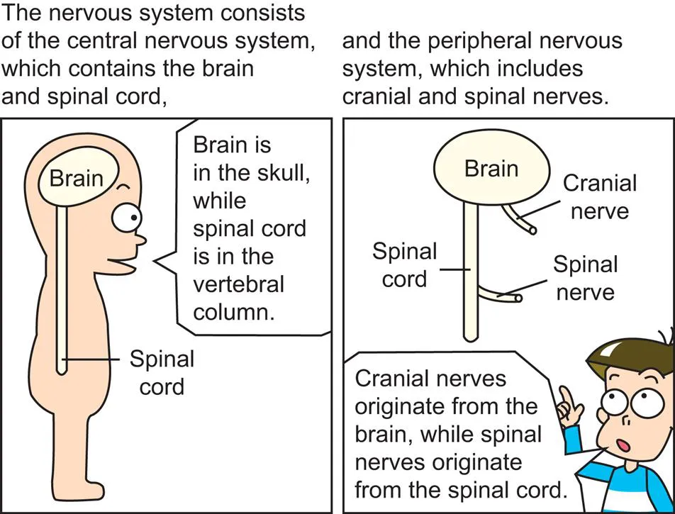
The nervous system is a complex network of nerves that carry impulses between the brain, spinal cord, and various parts of the body.
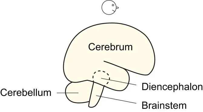
When the brain is viewed laterally, its three components are identifiable: cerebrum, cerebellum, and brainstem. The diencephalon is hidden by the cerebrum (cerebral hemisphere) (Fig. 1.11).
The blood supply, the cerebrospinal fluid flow

The basilar artery arises from the confluence of the two vertebral arteries at the junction between the pons and medulla oblongata. Branches of the basilar artery, named pontine arteries, feed the pons (Fig. 1.51).
The posterior inferior cerebellar artery branches off from the vertebral artery, while the anterior inferior cerebellar artery and superior cerebellar artery branch off from the basilar artery. This is because the basilar artery is on the pons (Fig. 1.54) which is right in front of the cerebellum (Figs. 1.44, 5.6).
There are three cerebral arteries as well as three cerebellar arteries on each side. The posterior cerebral artery is a terminal division of the basilar artery, while the middle and anterior cerebral arteries are two divisions of the internal carotid artery.
The “posterior” cerebral arteries and internal carotid arteries are connected by the “posterior” communicating arteries, while the bilateral “anterior” cerebral arteries are connected by the “anterior” communicating artery.
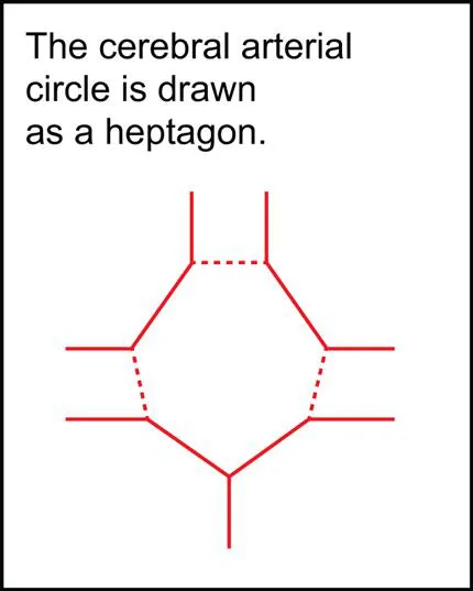
The cerebral arterial circle (circle of Willis) is composed of the posterior cerebral arteries, posterior communicating arteries, anterior cerebral arteries, and anterior communicating artery (Exactly, a short segment of internal carotid artery is included.) (Fig. 1.3). The circle is an anastomosis that guarantees blood supply to the cerebrum.

The anterior cerebral artery passes along the medial surface of the cerebral hemisphere anteriorly, superiorly, and then posteriorly. The middle cerebral artery emerges from the lateral sulcus to take charge of most of the lateral surface of the cerebral hemisphere (Fig. 1.23). The posterior cerebral artery passes posteriorly along its inferomedial surface (Figs. 1.6, 1.30).
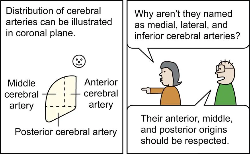
The anterior and middle cerebral arteries supply blood to the cerebral hemisphere above a certain horizontal plane; the posterior cerebral artery feeds the cerebral hemisphere below the plane (Fig. 1.5). In other words, the horizontal plane is a territorial border between the internal carotid artery and the vertebral artery (Fig. 1.3).
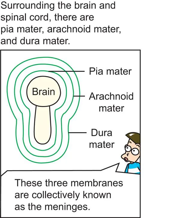
The meninges which cover the brain and spinal cord are like PAD. The meninges are composed of Pia, Arachnoid, and Dura maters.

The pia mater is adhesive to the brain (Figs. 1.14, 1.31) and spinal cord, the arachnoid (spider’s) mater is entangled like a spider’s web, and the dura mater is thick (Fig. 1.17).

The DURA mater reminds us of a DURAble mother.
The subarachnoid space of brain and spinal cord is an actual space containing cerebrospinal fluid ...
Table of contents
- Cover image
- Title page
- Table of Contents
- Copyright
- Preface
- Prologue
- Chapter 1. Morphology of the central nervous system
- Chapter 2. The somatic nerve, the autonomic nerve
- Chapter 3. The cranial nerve, the spinal nerve
- Chapter 4. Function of the brain
- Chapter 5. Development of the central nervous system
- Tables
- Other recommended readings
- Index
Frequently asked questions
Yes, you can cancel anytime from the Subscription tab in your account settings on the Perlego website. Your subscription will stay active until the end of your current billing period. Learn how to cancel your subscription
No, books cannot be downloaded as external files, such as PDFs, for use outside of Perlego. However, you can download books within the Perlego app for offline reading on mobile or tablet. Learn how to download books offline
Perlego offers two plans: Essential and Complete
- Essential is ideal for learners and professionals who enjoy exploring a wide range of subjects. Access the Essential Library with 800,000+ trusted titles and best-sellers across business, personal growth, and the humanities. Includes unlimited reading time and Standard Read Aloud voice.
- Complete: Perfect for advanced learners and researchers needing full, unrestricted access. Unlock 1.4M+ books across hundreds of subjects, including academic and specialized titles. The Complete Plan also includes advanced features like Premium Read Aloud and Research Assistant.
We are an online textbook subscription service, where you can get access to an entire online library for less than the price of a single book per month. With over 1 million books across 990+ topics, we’ve got you covered! Learn about our mission
Look out for the read-aloud symbol on your next book to see if you can listen to it. The read-aloud tool reads text aloud for you, highlighting the text as it is being read. You can pause it, speed it up and slow it down. Learn more about Read Aloud
Yes! You can use the Perlego app on both iOS and Android devices to read anytime, anywhere — even offline. Perfect for commutes or when you’re on the go.
Please note we cannot support devices running on iOS 13 and Android 7 or earlier. Learn more about using the app
Please note we cannot support devices running on iOS 13 and Android 7 or earlier. Learn more about using the app
Yes, you can access Visually Memorable Neuroanatomy for Beginners by Min Suk Chung,Beom Sun Chung in PDF and/or ePUB format, as well as other popular books in Biological Sciences & Human Anatomy & Physiology. We have over one million books available in our catalogue for you to explore.