Biomineralization is a natural process by which living organisms form minerals in association with organic biostructures to form hybrid biological materials such as bone, enamel, dentine and nacre among others. Scientists have researched the fundamentals of these processes and the unique structures and properties of the resulting mineralized tissues. Inspired by them, new biomaterials for tissue engineering and regenerative medicine have been developed in recent years.
Biomineralization and biomaterials: fundamentals and applications looks at the characteristics of these essential processes and natural materials and describes strategies and technologies to biomimetically design and produce biomaterials with improved biological performance.
- Provides a thorough overview of the biomineralization process
- Presents the most recent information on the natural process by which crystals in tissues form into inorganic structures such as bone, teeth, and other natural mineralized tissues
- Investigates methods for improving mineralization
- Explores new techniques that will help improve the biomimetic process
Trusted by 375,005 students
Access to over 1.5 million titles for a fair monthly price.
Design and engineering of biominerals and crystalline materials from the bottom up
J. Seto1,2; P.A. Romero31 University of California, San Francisco, CA, USA 2 École Normale Supérieure, Paris, France 3 University of California, Los Angeles, CA, USA
Abstract
Our awe about shapes and colors do not stop with our eyes; bound with imagination and curiosity, humanity has peered into these materials as symbols of strength, wisdom, as well as tranquility. We peer into its luster as our forefathers have done in the past and our children will in the future, to revel in what we see from its reflection in the present. We can rest to the solace and calm of its organization and its permanence will be present for generations to come.
Crystals come in all forms, sizes, and colors. Whether they be of the metallic, ionic, covalent, or molecular kind, the word “crystal” denotes order and stability. A material that is “crystalline” is composed of well-organized and “packed” constituent subunits. The crystals from a biological source intercalate this crystalline order with organic macromolecules to endow the assemblies with other physical characteristics to regulate form and function (i.e., size, hydrophobicity, electronegativity, and functional bonding). In this chapter, ionic crystals of biogenic origin as well as the biological mechanisms involved in the formation and growth of these diverse crystalline materials from Nature will be explored.
Keywords
Biomineralization
Mineral–organic interactions
Mesocrystalline
Hierarchical structuring
Directed evolution
Acknowledgments
This work would not have been possible without numerous collaborations with the following people: Profs. Fred H. Wilt, Peter Fratzl, Helmut Cölfen, Yong Chen, Stefan Habelitz, Malcolm Snead, Adam Abate, and Micheal Jewett. J. S. would like to acknowledge his family and Dr. Ozlem Sel for their unwavering support and continued presence in his professional life. P. R. acknowledges support from UCLA start-up funds.
1.1 Introduction
Humpty Dumpty sat on a wall,
Humpty Dumpty had a great fall,
All the King’s horses and all the King’s men,
[Finally re-crystallized] Humpty together again.
Unknown
We can search Nature’s abyss and find that we are, in fact, surrounded by crystalline-based materials. From the CaCO3 spines of sea urchins that populate the tide pools at the shoreline to the silica granules found in the body plans of sea sponges occupying the vast seas, these biogenic crystals have been sources of mineral that create geological landmarks such as cliffs and seafloors for eons of time. More impressively, each organism that produces these crystalline materials has its own “toolkit” of organic molecules that are tailored to create the structures and functions that allow these organisms to propagate onward into the future. The biological control and regulation involved in the formation and growth of these materials are also flexible enough to enable for additional revision as well as continued evolution, when making new biological mineral components.
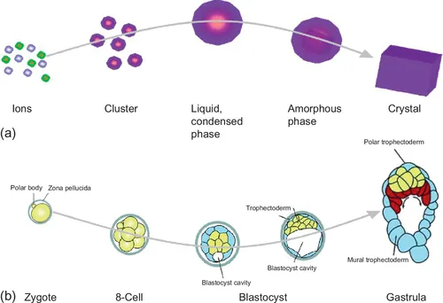
Some examples of the spatial, temporal control and precision involved in the regulation of crystal formation and growth can be witnessed through parallels with the basic premise of an embryo in biology. A mineral crystal forms from a nucleation event where a cation and anion pair bonds and creates a nuclei for crystal growth; an embryo forms from the union of female and male gamete cells and cellular developmental processes enable it to grow into an organism (Figure 1.1). In a similar fashion, the pathway a crystal nuclei will take will depend on its chemical and physical environments to determine its trajectory to becoming a fully fledged crystal with specific properties (i.e., polymorph, orientation, lattice spacings, and size) such as the path of crystal growth it will take and whether it will become one crystal polymorph versus another polymorph. Many of these environments can be controlled by biological macromolecules—proteins, lipids, and carbohydrates, as well as nucleic acids (Burke et al., 2000; Fantner et al., 2005; Vos et al., 2007; Knapp et al., 2012; Rao et al., 2014; Chow et al., 2014; Figure 1.2). By taking the simple scenario whereby the presence of biomolecules like nucleic acids, such as deoxyribonucleic acid (DNA), the carboxyl acid group (− COOH) can alter the bulk pH of solutions such that the local supersaturation of ion concentrations are slightly altered to induce a specific polymorph of a mineral (Xu et al., 2008; Figure 1.2). The presence of these molecules has been studied to a great degree and sheds light on how these molecules affect liquid–liquid as well as liquid–solid interfaces in crystal formation as well as the final crystalline product too (Li et al., 2012; Gebauer et al., 2008; Bewernitz et al., 2012; Gower, 2008; Burke et al., 2000; Lu et al., 2005).
Figure 1.1 Processes of crystallization and embryogenesis compared. (a) Ion pair bonding occurs to initiate nucleation and, subsequently, crystal growth. (b) Sperm and egg cells join to form a zygote that will undergo various stages of growth and development. Adapted from (a) Cölfen and Antonietti (2008) and (b) Beddington and Robertson (1999).
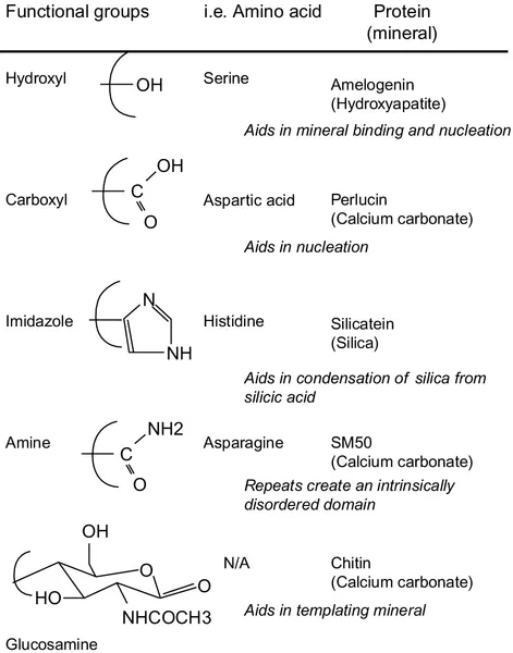
Figure 1.2 Typical functional groups found in biomineralization proteins and the amino acids that form them, known to alter solution properties during crystallization.
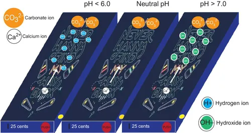
It may seem surprising to the reader that such “minor” alterations in the chemical environment, in this case a slight decrease of pH, can inflict such a dramatic outcome in crystallization. The pH, an indicator of the number of H+ ions in solution, more precisely related to the log of the concentration of H+ ions, inhibits the kinetics of the anion and thus affects the local supersaturations of the ion pairs involved in crystallization (Figure 1.3). By varying the pH in a crystallizing solution, whether it be lower (more H+ ions) or higher (more OH− groups), the cation and anion pair will be obstructed by the additional H+ or OH− ions in solution. As a result, the association and dissociation constants, respectively, will be affected above or below the pK of the cation and anion bonding pair (Figure 1.3). Thus, the degree and controls required to keep crystallization in biology in check necessitate regulatory schemes that can handle large amounts of ions as well as various potentials created by the ions.
Figure 1.3 Pinball schematic demonstrating the gamut of ions at low pH, neutral pH, and high pH conditions, showing problematic routes to mineralization in the low and high pH conditions.
In biology, regulation comes in the form of compartmentalization such that sensitive elements are localized and surrounded by protective membranes in order to create a buffer space separated from the surrounding cellular environment. Some examples of this compartmentalization in Nature can be witnessed in the formation of vesicles that are able to deliver extracellular payloads to specific intracellular organelles, as well as the redox processes occurring in the double membrane of the mitochondria, and finally, the membrane-mediated mineralization processes that occur in a growing sea urchin larvae spicule (Wilt et al., 2003), the growing tunic spicules in ascidians (Aizenberg et al., 2005), and the growing bone (Currey, 2005; Figures 1.4 and 1.5). Specifically, in the case of the growth of a sea urchin larvae spicule, the Ca2 + and
ions are transported into the primary mesenchymal cells during gastrulation along with matrix proteins into a syncytial space to enable nucleation of a precursor mineral that eventually grows into a single crystalline spicule made of CaCO3; subsequently, the same mechanism is adapted by the organism to form the large spines we often see at the beach (Wilt et al., 2003; Seto et al., 2004; Killian et al., 2009; Gong et al., 2012). In the formation and growth of the tunic spicules in ascidians, specific cells release localized silicatein proteins into an extracellular space that is surrounded by a proteinaceous membrane in order to convert silic acid into silica (Aizenberg et al., 2004, 2005). Beyond calcium carbonate and silica, a similar mechanism is utilized for hydroxyapatite in the initial mineralization and development of bone. In the formation of bone, osteoblasts, the cells that secrete minerals, and osteoclasts, the cells that resorb minerals, reach a secretion–resorption equilibrium such that osteoblasts secrete more minerals than are resorbed by osteoclasts (Cowin, 2001; Currey, 2002). The osteoblasts initially absorb minerals in the form of an amorphous precursor and, after intracellular processing, the minerals are secreted into a space called an osteoid that serves as the site of bone mineralization, mineral organization, and growth (Boskey, 1997). Whether it is an echinoderm, mollusk, or vertebrate, the mineralization mechanisms involved in all these diverse organisms share a common theme of membrane-mediated mineralization even though the components involved in each case, t...
Table of contents
Cover image
Title page
Table of Contents
Copyright
Contributors
Preface
Woodhead Publishing Series in Biomaterials
Part One: Fundamentals
Part Two: Applications
Index
Frequently asked questions
Yes, you can cancel anytime from the Subscription tab in your account settings on the Perlego website. Your subscription will stay active until the end of your current billing period. Learn how to cancel your subscription
No, books cannot be downloaded as external files, such as PDFs, for use outside of Perlego. However, you can download books within the Perlego app for offline reading on mobile or tablet. Learn how to download books offline
We are an online textbook subscription service, where you can get access to an entire online library for less than the price of a single book per month. With over 1.5 million books across 990+ topics, we’ve got you covered! Learn about our mission
Look out for the read-aloud symbol on your next book to see if you can listen to it. The read-aloud tool reads text aloud for you, highlighting the text as it is being read. You can pause it, speed it up and slow it down. Learn more about Read Aloud
Yes! You can use the Perlego app on both iOS and Android devices to read anytime, anywhere — even offline. Perfect for commutes or when you’re on the go. Please note we cannot support devices running on iOS 13 and Android 7 or earlier. Learn more about using the app
Yes, you can access Biomineralization and Biomaterials by Conrado Aparicio,Maria Pau Ginebra in PDF and/or ePUB format, as well as other popular books in Biological Sciences & Biotechnology in Medicine. We have over 1.5 million books available in our catalogue for you to explore.