eBook - ePub
Atlas of Hematopathology
Morphology, Immunophenotype, Cytogenetics, and Molecular Approaches
- 756 pages
- English
- ePUB (mobile friendly)
- Available on iOS & Android
eBook - ePub
Atlas of Hematopathology
Morphology, Immunophenotype, Cytogenetics, and Molecular Approaches
About this book
As the definitive diagnostic atlas of the diseases of the hematopoietic system, the Atlas of Hematopathology appeals to a wide range of people who are being trained in a variety of medical fields or practicing as non-hematopathologists, and therefore, are looking for a book which can provide information in a clear, focused format, with no excessive text or details. The atlas offers effective guidance in evaluating specimens from the lymph nodes, bone marrow, spleen, and peripheral blood, enabling clinicians to deliver more accurate and actionable pathology reports. Practicing physicians and those in pathology and hematology training also gain a better understanding of the nature of hematologic disorders and improve their diagnostic skills along the way. Taking a unique multi-disciplinary approach, the book covers conventional histopathology and cytopathology, as well as all important complementary diagnostic tests, such as immunophenotyping (immunohistochemical stains and flow cytometry), karyotyping, FISH and DNA/molecular studies. It offers concise textual and extensive visual coverage of both neoplastic and non-neoplastic hematology disorders, with the neoplastic hematology sections presented according to the most recent WHO classifications. There is also an introduction to the normal structures of hematopoietic tissues and the various multidisciplinary techniques. The atlas contains more than 900 high-quality color images that mirror the findings that fellows and clinicians encounter in practice. It provides information in a quick, simple and user-friendly manner, attracting those who are in training or are not considered experts in the field. Residents, fellows, practicing clinicians, and researchers in pathology, hematology, hematology/oncology, as well as graduate students in pathology and other clinicians workings in clinical hematology laboratories will all find it useful.
- Saves clinicians and researchers time in quickly accessing the very latest details on the diverse clinical and scientific aspects of hematopathology, as opposed to searching through thousands of journal articles
- For clinicians, fellows, and residents, correct diagnosis (and therefore correct treatment) of diseases depends on a strong understanding of the molecular basis for the disease – hematologists, pathologists, oncologists, and other clinicians will benefit from this clear, focused, annotated format
- Companion web site features over 900 images from the book!
Trusted by 375,005 students
Access to over 1.5 million titles for a fair monthly price.
Study more efficiently using our study tools.
Information
1
Structure and Function of Hematopoietic Tissues
Bone marrow is a mesenchymal-derived complex structure consisting of hematopoietic precursors and a complex microenvironment that facilitates the maintenance of hematopoietic stem cells (HSCs) and supports the differentiation and maturation of the progenitors (Figure 1.1). All differentiated hematopoietic cells including lymphocytes, erythrocytes, granulocytes, macrophages, and platelets are derived from HSCs (Figure 1.2).
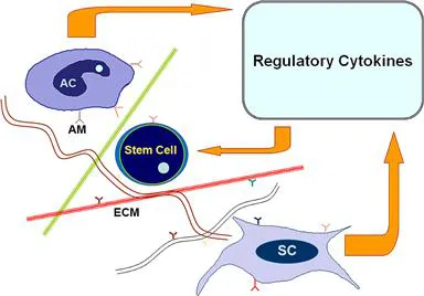
Figure 1.1 Cytokines released from accessory cells (AC) (e.g., macrophages, T-cells) and stromal cells have a regulatory effect on stem cells. The extracellular matrix (ECM) and adhesion molecules (AM) support cell–cell, cell–matrix, and cell–cytokine interactions.
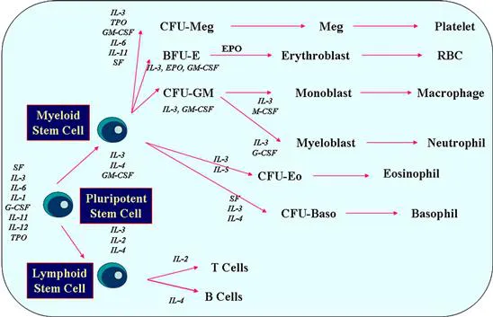
Figure 1.2 Current scheme of hematopoiesis demonstrating the differentiation of the multipotent stem cell to hematopoietic precursors and various levels of cytokine interaction.
The most primitive (pluripotent) HSCs express CD34 and are negative for CD38 and HLA-DR. These primitive cells, which include long-term repopulating stem cells, are also characterized by low-level expression of c-kit receptor (CD117) and absence of lineage specific maturation markers. There is a spectrum of heterogeneity in the bone marrow stem cell pool: a continuum of cells with decreasing capacity for self-renewal and increasing potential for differentiation. This trend is also associated with changes in immunophenotypic features. For example, the more mature committed stem cells, in addition to CD34, appear to express CD38 and/or HLA-DR. The pluripotent HSCs comprise about 1 per 20,000 of bone marrow cells, and only a small fraction of them are active, whereas the remaining majority are in a “resting” phase, on call for action when it is necessary. The HSCs reside in microenvironmental niches. These niches—which are composed of stromal cells, accessory cells (such as T lymphocytes and macrophages), components of extracellular matrix (Table 1.1), and various regulatory cytokines (Table 1.2) (Figure 1.1)—play an important role in the regulation of hematopoiesis and proliferation of the committed stem cells, leading to the production of huge numbers of progenitor cells and differentiated mature blood cells (Figure 1.2). Every day, an estimated 2.5 billion red cells, 2.5 billion platelets, and 1.0 billion granulocytes are produced per kilogram body weight in normal conditions.
Table 1.1 Major Components of Extracellular Matrix in Bone Marrow
| Type | Comments |
| Collagen (reticulin) | Consisting of various subtypes. Erythroid and myeloid precursors adhere to collagen types I and VI |
| Fibronectin | Attaches to early erythroid precursors and other hematopoietic and stromal cells |
| Hemonectin | Myeloid precursors adhere to laminin. Regulates leukocyte chemotaxis. |
| Proteoglycans | Components containing heparin sulfate, chondritin sulfate, and hyaluronic acid. Interact with laminin and type IV collagen and play a role in cytokine presentation and cell differentiation |
| Thrombospondin | Interacts with collagen, fibronectin, and CD36 |
Table 1.2 Regulatory Cytokines
| Cytokine | Primary Effect |
| GM-CSF1 | Granulocyte and macrophage colony formation, functional enhancement of mature forms |
| G-CSF2 | Granulocyte colony formation, functional enhancement of granulocytes |
| M-CSF (CSF-1)3 | Macrophage colony formation, functional enhancement of monocytes and macrophages |
| Erythropoietin (EPO) | Erythropoiesis, possible enhancement of megakaryocyte proliferation |
| Thrombopoietin (TPO) | Megakaryocyte proliferation, platelet production |
| Steel factor (c-kit ligand) | Stem cell and mast cell proliferation |
| Interleukin (IL)-1 | Promoter of hematopoiesis, inducer of other factors, B- and T-cell regulator, endogenous pyogen |
| IL-2 | T-cell growth factor, may inhibit G/M colony formation and erythropoiesis |
| IL-3 (Multi-CSF) | G/M colony formation, syngeneic effects on EPO, eosinophil, mast cell, and megakaryocyte colony formation |
| IL-4 | B-cell proliferation, IgE production |
| IL-5 | Eosinophil growth and B-cell differentiation |
| IL-6 | B-cell differentiation, synergistic effects on IL-1 |
| IL-7 | Development of B and T cell precursors |
| IL-8 | Granulocyte chemotactic factor |
| IL-9 | Growth of mast cells and T cells |
| IL-10 | Inhibitor of inflammatory and immune responses |
| IL-11 | Synergistic effects on growth of stem cells and megakaryocytes |
| IL-12 | Promoter of Th1 and suppressor of Th2 functions |
| IL-13 | B-cell proliferation, IgE production |
| IL-14 | High molecular weight B-cell growth factor |
| IL-15 | Activates T cells, neutrophils and macrophages |
| IL-16 | Chemotactic factor for helper T cells |
| IL-17 | Promotes T cell proliferation, pro-inflammatory activities |
| IL-18 | Activates T cells, neutrophils, and fibroblasts |
| IL-19 | Member of IL-10 family; transcriptional activator of IL-10 |
| IL-20 | Member of IL-10 family with epidermal function |
| IL-21 | Improves proliferation of T cells and B cells, and enhances natural killer (NK) cytotoxic activities |
| IL-22 | Member of IL-10 family; induces inflammatory responses |
| IL-23 | Activates autoimmune responses |
| IL-24 | Member of IL-10 family; tumor suppressor molecule |
| IL-25 | Capable of amplifying allergic inflammation |
| IL-26 | Member of IL-10 family; plays a role in mucosal and cutaneous immunity |
| TGF4-β | Suppresses BFU-E, CFU-S, and HPP-CFC |
| Interferons | Suppress BFU-E, CFU-GEMM, and CFU-GM |
| TNF5-α and -β | Suppress BFU-E, CFU-GEMM, and CFU-GM |
| PGE6-1 and -2 | Suppress GFU-GM, GFU-G, and GFU-M |
| Lactoferrin | Suppresses release of IL-1 |
1Granulocyte and macrophage colony-stimulating factor
2Granulocyte colony-stimulating factor
3Macrophage colony-stimulating factor
4Transforming growth factor
5Tumor necrosis factor
6Prostaglandin E
Bone marrow stromal cells are derived from pluripotent stromal stem cells. In other words, two separate and distinct pluripotent stem cells are simultaneously at work in bone marrow: hematopoietic and stromal. These two systems not only coexist but closely interact with each other. Stromal cells are composed of a heterogeneous cell population including adipocytes, fibroblast-like cells, endothelial cells, and osteoblasts. They produce a number of cytokines and a group of proteins that are involved in facilitating cell–cell interactions and presenting the cytokines and growth factors to the hematopoietic progenitor cells. Stromal cells with their extracellular matrix make a fibrovascular mesh environment to home and support the hematopoietic precursors. The thin-walled venous sinuses are the most prominent vascular spaces in the bone marrow. They consist of an inner layer of endothelial cells supported by an outer layer of fibroblast-like (parasinal, adventitial) stromal cells. They receive blood from the branches of the nutrient artery and periosteal capillary network. The nutrient artery penetrates the bony shaft, branches into the bone marrow cavity, and forms capillary...
Table of contents
- Cover image
- Title page
- Table of Contents
- Copyright
- Preface
- Acknowledgments
- 1. Structure and Function of Hematopoietic Tissues
- 2. Principles of Immunophenotyping
- 3. Principles of Cytogenetics
- 4. Principles of Molecular Techniques
- 5. Morphology of Abnormal Bone Marrow
- 6. Reactive Lymphadenopathies
- 7. Bone Marrow Aplasia
- 8. Myelodysplastic Syndromes/Neoplasms—Overview
- 9. Myelodysplastic Syndromes/Neoplasms—Classification
- 10. Myeloproliferative Neoplasms—Overview
- 11. Chronic Myelogenous Leukemia
- 12. Myeloproliferative Neoplasms Associated with JAK2 Mutation
- 13. Chronic Neutrophilic and Chronic Eosinophilic Leukemias
- 14. Mastocytosis
- 15. Myelodysplastic/Myeloproliferative Neoplasms
- 16. Hematologic Neoplasms Associated with Eosinophilia and PDGFRA, PDGFRB, or FGFR1 Rearrangement
- 17. Acute Myeloid Leukemia—Overview
- 18. Acute Myeloid Leukemias with Recurrent Genetic Abnormalities
- 19. Acute Myeloid Leukemia with Myelodysplasia-Related Changes
- 20. Therapy-Related Myeloid Neoplasms
- 21. Acute Myeloid Leukemia, Not Otherwise Specified
- 22. Myeloid Proliferations Related to Down Syndrome
- 23. Lymphoblastic Neoplasms—B-Lymphoblastic Leukemia/Lymphoma
- 24. Lymphoblastic Neoplasms—T-Lymphoblastic Leukemia/Lymphoma
- 25. Acute Leukemias of Ambiguous Lineage
- 26. Mature B-Cell Neoplasms—Overview
- 27. Chronic Lymphocytic Leukemia/Small Lymphocytic Lymphoma
- 28. B-Cell Prolymphocytic Leukemia
- 29. Lymphoplasmacytic Lymphoma/Waldenström’s Macroglobulinemia
- 30. Hairy Cell Leukemia
- 31. Splenic Marginal Zone Lymphoma
- 32. Nodal Marginal Zone Lymphoma
- 33. Extranodal Marginal Zone Lymphoma of Mucosa-Associated Lymphoid Tissue (MALT Lymphoma)
- 34. Follicular Lymphoma
- 35. Mantle Cell Lymphoma
- 36. Diffuse Large B-Cell Lymphoma
- 37. Diffuse Large B-Cell Lymphoma Subtypes
- 38. Other Lymphomas of Large B Cells
- 39. Burkitt Lymphoma
- 40. Primary Cutaneous B-Cell Lymphomas
- 41. Plasma Cell Neoplasms
- 42. Mature T- and NK-Cell Neoplasms—Overview
- 43. Large Granular Lymphocytic Neoplasms and Related Disorders
- 44. T-Cell Prolymphocytic Leukemia
- 45. Adult T-Cell Leukemia/Lymphoma
- 46. Hepatosplenic T-Cell Lymphoma
- 47. Enteropathy-Associated T-Cell Lymphoma
- 48. Mycosis Fungoides and Sézary Syndrome
- 49. Other Primary Cutaneous T-Cell Lymphoproliferative Disorders
- 50. Angioimmunoblastic T-Cell Lymphoma
- 51. Anaplastic Large Cell Lymphomas
- 52. Peripheral T-Cell Lymphoma, Not Otherwise Specified
- 53. Nodular Lymphocyte Predominant Hodgkin Lymphoma
- 54. Classical Hodgkin Lymphoma
- 55. Immunodeficiency Disorders
- 56. Iatrogenic Immunodeficiency-Associated Lymphoproliferative Disorders
- 57. Lymphocytopenia and Lymphocytosis
- 58. Histiocytic Disorders
- 59. Disorders of Dendritic Cells
- 60. Granulocytic Disorders
- 61. Disorders of Red Blood Cells—Anemias
- 62. Disorders of Megakaryocytes and Platelets
- 63. Post-Therapy Changes
- Index
Frequently asked questions
Yes, you can cancel anytime from the Subscription tab in your account settings on the Perlego website. Your subscription will stay active until the end of your current billing period. Learn how to cancel your subscription
No, books cannot be downloaded as external files, such as PDFs, for use outside of Perlego. However, you can download books within the Perlego app for offline reading on mobile or tablet. Learn how to download books offline
We are an online textbook subscription service, where you can get access to an entire online library for less than the price of a single book per month. With over 1.5 million books across 990+ topics, we’ve got you covered! Learn about our mission
Look out for the read-aloud symbol on your next book to see if you can listen to it. The read-aloud tool reads text aloud for you, highlighting the text as it is being read. You can pause it, speed it up and slow it down. Learn more about Read Aloud
Yes! You can use the Perlego app on both iOS and Android devices to read anytime, anywhere — even offline. Perfect for commutes or when you’re on the go.
Please note we cannot support devices running on iOS 13 and Android 7 or earlier. Learn more about using the app
Please note we cannot support devices running on iOS 13 and Android 7 or earlier. Learn more about using the app
Yes, you can access Atlas of Hematopathology by Faramarz Naeim in PDF and/or ePUB format, as well as other popular books in Biological Sciences & Oncology. We have over 1.5 million books available in our catalogue for you to explore.