Neuromuscular Function and Disorders focuses on the various processes underlying disordered neuromuscular function. Topics covered include the nature of membrane defects in myotonia and familial periodic paralysis; the disorder of neuromuscular transmission responsible for myasthenia gravis and the various pseudo-myasthenic syndromes; and the disorders of Schwann cell function which cause demyelination. This book is comprised of 28 chapters divided into two sections and begins with a discussion on the normal anatomy and physiology of peripheral nerve and muscle. Included in the first section are descriptions of the ionic mechanisms responsible for the resting and action potentials of nerve and muscle; the sequential stages in neuromuscular transmission; excitation-contraction coupling; the sliding filament mechanism of myofibrillar shortening; and the morphological and functional properties of motor units. The neurophysiology of exercise and muscle fatigue is also considered, along with the nature of the trophic influences exerted by the motoneuron and muscle fiber upon each other. The second half of the book deals entirely with various diseases of peripheral nerve and muscle, together with diagnostic procedures and therapeutic management. A consistent theme in this section is the recognition of neural abnormalities in diseases hitherto considered as primary disorders of the muscle fiber. This monograph should be of value to neurologists, medical students, research workers, and students and research scientists in physiology, zoology, pharmacology, kinesiology, and physical education.
Chapter 8: TROPHIC INTERACTIONS OF NERVE AND MUSCLE: DENERVATION AND REINNERVATION
Chapter 9: USE AND DISUSE
Chapter 10: TROPHIC INFLUENCE OF MUSCLE ON NERVE
Chapter 11: MUSCLE GROWTH
Chapter 12: AGEING
Chapter 1
THE MUSCLE FIBRE
Publisher Summary
This chapter discusses the structure of muscle fiber. Voluntary” muscles are composed of two types of muscle fibers. By far the most common are those fibers that make up nearly the entire bulk of the muscle and are described as extrafusal. This term distinguishes them from the much smaller muscle fibers that are found inside the muscle spindles. Because of their location, these smaller muscle fibers are referred to as intrafusal. At birth, human fibers are slender, measuring about 10–20 μm in diameter. Throughout childhood and particularly during the growth spurt in puberty, the sizes increase and eventually attain adult dimensions with most diameters in the 40–80 μm range. The membrane of the muscle fiber is termed the sarcolemma and is some 75 A thick. Just outside the sarcolemma and clearly visible in electronmicrographs is the basement membrane which is composed of proteins and polysaccharides. Each muscle fiber contains numerous nuclei which are dispersed along the inner surface of the sarcolemma, particularly in the region of the motor end-plate. The most obvious structures within the muscle fiber are the myofibrils, which are the units responsible for contraction and relaxation of the fiber.
The ‘voluntary’ muscles are composed of muscle fibres, of which two types are found. By far the commonest are those fibres which make up nearly all the bulk of the muscle and are described as extrafusal. This term distinguishes them from the much smaller muscle fibres which are found inside the muscle spindles; because of their location these last fibres are referred to as intrafusal. The special structure and function of the intrafusal fibres are of considerable interest and have been reviewed elsewhere (Matthews, 1972). This book will deal only with the extrafusal muscle fibres, however, for it is these fibres about which most is known in disease and it is their ineffectiveness which is ultimately responsible for the cardinal symptom of weakness.
NUMBERS AND SIZES OF MUSCLE FIBRES
Because of the time-consuming nature of the task involved, there are very few values for the numbers of extrafusal fibres in human voluntary muscles. On the basis of fibre counts in samples taken from cadaveric tissue, Feinstein et al. (1955) made the estimates set out in Table 1.1.
TABLE 1.1
Number of Muscle Fibres in Various Human Muscles (results given to nearest 50)
Muscle
No. of muscle fibres
Author
First lumbrical
10 250*
Feinstein et al. (1955)
External rectus
27 000
Feinstein et al. (1955)
Platysma
27 000
Feinstein et al. (1955)
First dorsal interosseous
40 500
Feinstein et al. (1955)
Sartorius
128 150*
MacCallum (1898)
Brachioradialis
129 200*
Feinstein et al. (1955)
Tibialis anterior
271 350
Feinstein et al. (1955)
Medial gastrocnemius
1 033 000
Feinstein et al. (1955)
*Average values
The long gestation period in man allows sufficient time for the precursors of the muscle fibres to have finished dividing and to have formed the adult complement of fibres before birth (MacCallum, 1898). In species with a shorter gestation period the number of fibres continues to increase in the neonatal period. At birth the human fibres are slender, measuring about 10–20 μm in diameter. Throughout childhood, and particularly during the growth spurt in puberty, the sizes increase and eventually attain adult dimensions, with most diameters in the 40–80 μm range (see Figure 1 of Brooke and Engel, 1969). So far as length is concerned, it is assumed that individual fibres run from one end of the muscle to the other; in the human sartorius muscle such fibres may not be functionally continuous; certainly the thigh muscles possess more than one innervation zone and it is possible that long fibres may be composed of two or more shorter fibres arranged end-to-end.
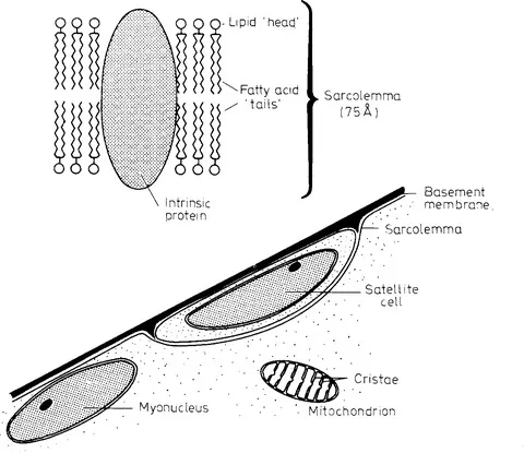
Figure 1.1 (Upper) Structure of the sarcolemma, showing the double layer of lipid molecules and an ‘intrinsic’ protein (see text). (Lower) Membranes, nuclei and organelles seen at the surface of a muscle fibre; the mitochondrion would measure about 1.5 μm in its long diameter
STRUCTURE OF THE MUSCLE FIBRE
Sarcolemma (plasmalemma)
All living cells are bounded by a membrane; that of the muscle fibre is termed the sarcolemma and is some 75
thick. The sarcolemma resembles the membrane of the nerve fibre in possessing the special property of excitability, enabling electrical impulses to be transmitted down the length of the cell (see page 20). In the region of the innervation zone (motor end-plate) the sarcolemma is highly convoluted, due to the presence of junctional folds (page 27). Elsewhere along the surface of the fibre the membrane displays much shallower folds; these result from slackness of the membrane when the fibre is in its resting or contracted states and they disappear if the fibre is passively stretched. There are also numerous very small in-pocketings of membrane, the caveolae, which are connected to the surface membrane by narrow necks; their function is uncertain though they can also act as reserve sources of membrane during stretching of the fibre (Dulhunty and Franzini-Armstrong, 1975). Biochemical and ultrastructural investigations indicate that the sarcolemma is largely composed of lipid molecules arranged perpendicularly to the surface of the fibre and forming two layers (Figure 1.1, upper; see also Capaldi, 1974). The hydrophilic ‘heads’ of the lipid molecules form the internal and external surfaces of the membrane while the hydrophobic ‘tails’ make up the interior of the membrane. The membrane also contains proteins, of which two types are generally recognized; these are referred to as extrinsic and intrinsic. Extrinsic proteins are only attached at the internal or external surfaces of the membrane and can be dislodged relatively easily by chemical means. In contrast, intrinsic proteins penetrate the full thickness of the membrane and are difficult to remove. Among the special proteins known to be localized in the sarcolemma are (a) transport systems for sugars, lipids and, amino acids; (b) kinases for phosphorylating various membrane proteins; (c) adenylate cyclase, responsible for synthesizing cyclic AMP; (d) ATPase enzymes for pumping cations across the membrane (one type for sodium and potassium and another for calcium and magnesium); and (e) carrier molecules (ionophores), each of which can select one species of ion (sodium, potassium or chloride) and transfer it across the membrane (see page 24). In the end-plate region the sarcolemma also contains two special proteins which combine with acetylcholine; these are the acetylcholine receptor and the hydrolytic enzyme, acetylcholinesterase. Since much of the membrane lipid has a melting point below body-temperature the sarcolemma would be expected to have fluid properties. By labelling a spot of membrane with a fluorescent dye and then observing its enlargement under the microscope Fambrough et al. (1974) were able to show that this was indeed the case.
Basement membrane
Just outside the sarcolemma and clearly visible in electronmicrographs is the basement membrane (Figure 1.1, lower). This membrane is about 500
thick and is composed of proteins and polysaccharides. It is probable that the basement membrane provides a special ionic milieu for the muscle fibre by restricting the further diffusion of electrolytes once these have crossed the sarcolemma. In addition the basement membrane helps to maintain the shape of the muscle fibre by providing external support. If the muscle fibre is damaged the membrane may be spared and can then guide the regenerating myoblasts in the formation of a new fibre.
Nuclei
Each muscle fibre contains numerous nuclei (myonuclei) which, in health, are dispersed along the inner surface of the sarcolemma, particularly in the region of the motor end-plate. The nucleus is bounded by two membranes, the outer one of which may join the sarcoplasmic reticulum (Figure 1.1, lower).
Indistinguishable from the myonuclei with the light microscope are the nuclei of the satellite cells. These cells probably account for less than 1 per cent of the muscle fibre nuclei in the adult. They can only be differentiated from the myonuclei by the electronmicroscope, which reveals the presence of twin membranes separating the cytoplasm of the satellite cell from that of the muscle fibre. The satellite cells are of particular importance for the regeneration of muscle following disease or injury (page 100).
Myofibrils
The most obvious structures within the muscle fibre are the myofibrils, which are the units responsible for contraction and relaxation of the fibre (Figure 1.2). Each myofibril is about 1 μm in diameter and, even with the light microscope, can be seen to have a banded, or striated, appearance. The ‘dark’ and ‘light’ striations are termed the A- and I-bands respectively; in the relaxed muscle fibre the centre of the A-band has a light region, the H-zone, which is itself bisected by a ‘dark’ M-line. In the centre of the I-band is a dark Z-line (Z-disc). The striations are caused by the fact that the part of the myofibril in the A-band has a refractive index which is substantially higher than that of the fluid medium, or sarcoplasm, in which the myofibrils are embedded. The pattern of one dark band followed by one light band is repeated every 2.2 μm (measured with the muscle in the relaxed state), the segment between two successive Z-lines being termed a sarcomere. All the myofibrils within a healthy muscle fibre are in register, such that the light and dark bands of one myofibril are adjacent to the cor...
Table of contents
Cover image
Title page
Table of Contents
Dedication
Copyright
INTRODUCTION
SYMBOLS, UNITS OF MEASUREMENT AND ABBREVIATIONS
PART 1: MUSCLE FIBRES AND MOTONEURONES
PART 2: DISORDERS OF MUSCLE AND NERVE
APPENDICES
REFERENCES
INDEX
Frequently asked questions
Yes, you can cancel anytime from the Subscription tab in your account settings on the Perlego website. Your subscription will stay active until the end of your current billing period. Learn how to cancel your subscription
No, books cannot be downloaded as external files, such as PDFs, for use outside of Perlego. However, you can download books within the Perlego app for offline reading on mobile or tablet. Learn how to download books offline
Perlego offers two plans: Essential and Complete
Essential is ideal for learners and professionals who enjoy exploring a wide range of subjects. Access the Essential Library with 800,000+ trusted titles and best-sellers across business, personal growth, and the humanities. Includes unlimited reading time and Standard Read Aloud voice.
Complete: Perfect for advanced learners and researchers needing full, unrestricted access. Unlock 1.4M+ books across hundreds of subjects, including academic and specialized titles. The Complete Plan also includes advanced features like Premium Read Aloud and Research Assistant.
Both plans are available with monthly, semester, or annual billing cycles.
We are an online textbook subscription service, where you can get access to an entire online library for less than the price of a single book per month. With over 1 million books across 990+ topics, we’ve got you covered! Learn about our mission
Look out for the read-aloud symbol on your next book to see if you can listen to it. The read-aloud tool reads text aloud for you, highlighting the text as it is being read. You can pause it, speed it up and slow it down. Learn more about Read Aloud
Yes! You can use the Perlego app on both iOS and Android devices to read anytime, anywhere — even offline. Perfect for commutes or when you’re on the go. Please note we cannot support devices running on iOS 13 and Android 7 or earlier. Learn more about using the app
Yes, you can access Neuromuscular Function and Disorders by Alan J. McComas in PDF and/or ePUB format, as well as other popular books in Medicine & Diseases & Allergies. We have over one million books available in our catalogue for you to explore.