Hemocompatibility of Biomaterials for Clinical Applications: Blood-Biomaterials Interactions summarizes the state-of-the-art on this important subject. The first part of the book reviews the latest research on blood composition and response, mechanisms of coagulation, test standards and methods. Next, the book assesses techniques for modifying biomaterial surfaces and developing coatings to improve hemocompatibility. In the final sections, users will find discussions on ways to improve the hemocompatibility of particular classes of biomaterials and a review of methods for improving medical devices.
- Provides comprehensive information on the fundamentals of hemocompatibility and new technologies
- Combines research in the biomaterials field in a digestible format for clinical applications
- Provides a complete overview biomaterials in current use and test methods
Trusted by 375,005 students
Access to over 1.5 million titles for a fair monthly price.
Fundamentals and testing methods for the hemocompatiblity of biomaterials
1
Contact activation by the intrinsic pathway of blood plasma coagulation
Y. Yan; L.-C. Xu; E.A. Vogler1; C.A. Siedlecki The Pennsylvania State University, Hershey, PA, United States 1 Deceased April 3, 2017
Abstract
It is well known that the biomaterial surfaces that comprise biomedical devices will initiate blood coagulation. This can occur through the adhesion and activation of platelets, but also through the activation of the proteins of the intrinsic coagulation cascade. Nominally, this involves activation of the zymogen FXII (Hageman Factor) which in turn activates FXI, prekallikrein and high-molecular-weight kininogen to form an activation complex. The end result of this sequence of zymogen/enzyme conversion steps is the production of thrombin, the conversion of fibrinogen to fibrin and the development of fibrin strands. In this chapter, we discuss the proteins of the intrinsic pathway, the traditional view of contact activation, and finally alternative ideas about the activation of this important pathway. Notably, we attempt to reconcile the inconsistencies between protein adsorption and function at biomaterial surfaces and the traditional views of contact activation.
Biomaterial surfaces initiate blood coagulation. This poses a significant challenge to the development of blood-contacting devices used in human beings and especially for the implementation of cardiovascular devices. The performance of medical products such as catheters, blood vessel grafts, vascular stents, extracorporeal oxygenator membranes, and left ventricular assistant device are significantly impaired by thrombosis problems [1]. A thrombus is composed of cross-linked fibrin clots and aggregated platelets connected by fibrinogen. When thrombi come off the site as emboli and travel through the blood stream, they may block small vessels downstream leading to stroke or tissue death. With the increasing demand for cardiovascular healthcare worldwide, it is imperative to understand the mechanisms leading to thrombus formation on biomaterial surfaces in order to develop strategies for improved hemocompatibility.
Blood coagulation can be potentiated by plasma-phase coagulation and/or platelet-mediated reactions [2]. When a biomaterial is implanted and/or blood vessel is injured, a variety of biological responses are initiated triggering processes of platelet activation/aggregation and plasma coagulation. The mechanism of platelet aggregation involves the adhesion, activation, and aggregation of platelets at the site of injured wall or on implanted biomaterial surfaces [3]. Plasma coagulation is a series of zymogen-to-enzyme conversions that occur as a cascade, in which the enzyme produced in one reaction catalyzes the next reaction. It penultimately produces thrombin (FIIa), a powerful serine protease, which hydrolyzes fibrinogen into fibrin monomers [2]. Fibrin monomers then oligomerize and cross-link into a mesh on and between aggregated platelets to form a mechanically stabilized aggregate. Platelet activation/aggregation and blood plasma coagulation are seemingly independent events, but in reality, there are strong interactions between the processes. The surface membrane of activated platelets promotes certain coagulation reactions, and FIIa itself is a potent platelet activator [3].
The plasma coagulation cascade is usually divided into two branches, termed the extrinsic and intrinsic pathways, that are dependent on the initial trigger. The extrinsic pathway is primarily responsible for hemostasis following vascular injury. Tissue factor (TF) embedded in the vessel walls is exposed after blood vessel injury and binds activated plasma coagulation factor VII (FVIIa). This surface-bound complex potentiates the blood coagulation by activating FIX and FX to FIXa and FXa, respectively [3]. Understanding of the intrinsic pathway dates back to the pioneering work of Oscar Ratnoff who discovered the indispensable role of Hageman factor (HF, otherwise known as factor XII, FXII) in promoting glass-induced blood coagulation in the 1950s [4]. The intrinsic pathway is initiated by biomaterial surface contact-induced activation of FXII to FXIIa (a.k.a. contact activation). This leads to the formation of a pathological thrombus in response to blood contact with foreign surfaces. Contact activation has been implicated as one of the causes for poor hemocompatibility of cardiovascular biomaterials. Both the intrinsic and extrinsic pathways eventually merge into the common pathway, leading to the formation of thrombin (FIIa), followed by fibrin monomer formation and oligomerization (Fig. 1.1). Note that plasma coagulation also involves feedforward or feedback loops in which an enzyme produced in one step acts as an inhibitor or activator in other reactions, which are not shown in Fig. 1.1 for simplicity.
Fig. 1.1 Simplified diagrams of plasma coagulation cascade with emphasis on the intersections of intrinsic and extrinsic pathways. Mediators, cofactors, inhibitors, and amplification loops are not shown for simplicity [2].
It has been widely accepted that FXII activation is specific only to negatively charged surfaces or compounds such as kaolin, glass, and ellagic acid. Recent studies revealed that FXII is activated in neat buffer solution at equal efficiency by contact with either hydrophobic or hydrophilic surfaces [5]. In addition, several “natural” surfaces have been identified to activate FXII in vivo, such as platelet polyphosphate, microparticles (MPs) derived from platelets and erythrocytes, RNA, amyloid β aggregates, misfolded proteins, collagen, and mast cell heparin [6–13]. Evidence shows that the presence of few platelets was enough to propagate coagulation substantially if contact activation was simultaneously initiated, indicating that plasma contact activation and platelet adhesion had a strong synergetic effect on thrombosis in blood-contacting materials [14].
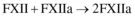
It is proposed that extent of FXII activation is related to the plasma coagulation. Previous studies suggested that activated FXII can be produced via at least three different biochemical pathways: autoactivation produces FXIIa by contact of FXII with an activating surface (
), reciprocal activation of FXII and prekallikrein (PK) (FXII is enzymatically cleaved by kallikrein presumably generated by FXIIa-mediated hydrolysis of PK), and autohydrolysis of FXII by (
) [15]. In this chapter, we will review the current understanding of FXII contact activation and recent insights into the process and introduce some new perspectives on FXII activation.
The efficiency of FXII activation is conventionally measured by functional assays in terms of enzymatic activities of FXII-derived products, generally referred to as procoagulant activity and amidolytic activity. Procoagulant activity is measured by simple plasma clotting assays (related to whole blood clotting tests) for the potential of FXII-derived products to induce clot formation (procoagulant activity). Procoagulant activity can be determined by comparing with a standard curve relating exogenously added αFXIIa, synthetic material surfaces, or other known activators to the clotting time. Amidolytic activity is assessed using various chromogenic assays as the ability to cleave a commercial chromogenic peptide in response to the presence of enzyme. Quantification of amidolytic activity is carried out by monitoring the color development due to proteolytic cleavage of the chromogen by FXIIa or other FXII-derived products. Both assays measure the net activity of the total FXII-derived proteins following activation without differentiating various proteins [16].
1.2 Structural a...
Table of contents
Cover image
Title page
Table of Contents
Copyright
Contributors
Part One: Fundamentals and testing methods for the hemocompatiblity of biomaterials
Part Two: Improving the hemocompatibility of biomaterial surfaces
Part Three: Improving the hemocompatibility of types of biomaterial
Part Four: Biomedical applications of hemocompatible biomaterials
Index
Frequently asked questions
Yes, you can cancel anytime from the Subscription tab in your account settings on the Perlego website. Your subscription will stay active until the end of your current billing period. Learn how to cancel your subscription
No, books cannot be downloaded as external files, such as PDFs, for use outside of Perlego. However, you can download books within the Perlego app for offline reading on mobile or tablet. Learn how to download books offline
Perlego offers two plans: Essential and Complete
Essential is ideal for learners and professionals who enjoy exploring a wide range of subjects. Access the Essential Library with 800,000+ trusted titles and best-sellers across business, personal growth, and the humanities. Includes unlimited reading time and Standard Read Aloud voice.
Complete: Perfect for advanced learners and researchers needing full, unrestricted access. Unlock 1.5M+ books across hundreds of subjects, including academic and specialized titles. The Complete Plan also includes advanced features like Premium Read Aloud and Research Assistant.
Both plans are available with monthly, semester, or annual billing cycles.
We are an online textbook subscription service, where you can get access to an entire online library for less than the price of a single book per month. With over 1.5 million books across 990+ topics, we’ve got you covered! Learn about our mission
Look out for the read-aloud symbol on your next book to see if you can listen to it. The read-aloud tool reads text aloud for you, highlighting the text as it is being read. You can pause it, speed it up and slow it down. Learn more about Read Aloud
Yes! You can use the Perlego app on both iOS and Android devices to read anytime, anywhere — even offline. Perfect for commutes or when you’re on the go. Please note we cannot support devices running on iOS 13 and Android 7 or earlier. Learn more about using the app
Yes, you can access Hemocompatibility of Biomaterials for Clinical Applications by Christopher Siedlecki in PDF and/or ePUB format, as well as other popular books in Biological Sciences & Biotechnology. We have over 1.5 million books available in our catalogue for you to explore.