- 344 pages
- English
- ePUB (mobile friendly)
- Available on iOS & Android
eBook - ePub
About this book
Arteriovenous and Cavernous Malformations, Volume 143, is the latest addition in the ongoing HCN series, an evidence-based compendium which addresses both the scientific and clinical aspects of this unique disease process. The volume covers didactic aspects, such as the epidemiology, etiology, and diagnosis of AVMs, while also providing expert clinical information on the management and treatment of these lesions.In addition, it provides coverage of modern-day advances in the genetics of cavernous malformations, as well as discussion regarding future open research questions. Readers from the laboratory bench to the bedside can expect a broad, yet objective, review of this pathology, with updates from the latest scientific literature and data supporting current practices.- Offers an evidence-based focus with coverage of both the scientific and clinical aspects of cavernous malformations- Addresses epidemiology, etiology, diagnosis, and genetics- Contains clinical insights regarding indications for surgery, surgical techniques, outcomes, and prognostic factors drawn from the authors' extensive experiences- Edited work with chapters authored by leaders in the field from around the globe – the broadest, most expert coverage available
Tools to learn more effectively

Saving Books

Keyword Search

Annotating Text

Listen to it instead
Information
Part I
Arteriovenous Malformations
Section 1
Cerebral arteriovenous malformations
Chapter 1
Epidemiology, genetics, pathophysiology, and prognostic classifications of cerebral arteriovenous malformations
Alp Ozpinar1,*; Gustavo Mendez2; Adib A. Abla3 1 Department of Neurological Surgery, University of Pittsburgh Medical Center, Pittsburgh, PA, USA
2 Department of General Surgery, Rush University Medical Center, Chicago, IL, USA
3 Department of Neurological Surgery, University of Arkansas for Medical Sciences, Little Rock, AR, USA
* Correspondence to: Alp Ozpinar, MD, Department of Neurological Surgery, University of Pittsburgh Medical Center, Suite B-400, 200 Lothrop Street, Pittsburgh PA, USA. Tel: +1-412-647-3685 email address: [email protected]
Abstract
Arteriovenous malformations (AVMs) are vascular deformities involving fistula formation of arterial to venous structures without an intervening capillary bed. Such anomalies can prove fatal as the high arterial flow can disrupt the integrity of venous walls, thus leading to dangerous sequelae such as hemorrhage. Diagnosis of these lesions in the central nervous system can often prove challenging as intracranial AVMs represent a heterogeneous vascular pathology with various presentations and symptomatology. The literature suggests that most brain AVMs (bAVMs) are identified following evaluation of the etiology of acute cerebral hemorrhage, or incidentally on imaging associated with seizure or headache workup. Given the low incidence of this disease, most of the data accrued on this pathology comes from single-center experiences. This chapter aims to distill the most important information from these studies as well as examine meta-analyses on bAVMs in order to provide a comprehensive introduction into the natural history, classification, genetic underpinnings of disease, and proposed pathophysiology. While there is yet much to be elucidated about AVMs of the central nervous system, we aim to provide an overview of bAVM etiology, classification, genetics, and pathophysiology inherent to the disease process.
Keywords
brain arteriovenous malformation; classification; pathophysiology
Introduction
Vascular disease entities manifest in various forms, and are particularly dangerous when high-flow arterial systems erroneously communicate directly with venous systems. Arteriovenous malformations (AVMs) are vascular deformities involving fistula formation of arterial to venous structures without an intervening capillary bed. Such anomalies can prove fatal as the high arterial flow can disrupt the integrity of venous walls, thus leading to sequelae such as hemorrhage. The central nervous system (CNS) is particularly sensitive to such insults as intracranial hemorrhage secondary to AVM rupture can prove profoundly disabling or deadly. Diagnosis of these lesions can prove challenging, as intracranial AVMs represent a heterogeneous vascular pathology with various presentations and symptomatology, as well as a wide variation in severity of the effect of an AVM on a patient's overall well-being. The literature suggests that most brain AVMs (bAVMs) are identified following evaluation of the etiology of acute cerebral hemorrhage, or incidentally on imaging associated with seizure or headache workup (Hofmeister et al., 2000; Conger et al., 2015). As reinforced by Conger et al. (2015), subsequent to detection of the AVM, history and physical examination coupled with critical imaging, including computed tomography (CT), magnetic resonance imaging (MRI), and catheter angiogram, allow neurosurgeons to form a clinical picture of the AVM patient as well as visualize the structure and hemodynamics of the vascular malformation (Conger et al., 2015).
To date, bAVM data have been derived from single-center experiences. From epidemiological studies, the incidence of AVMs ranges from 1.12 to 1.42 cases per 100 000 person-years, with 38–68% of new cases presenting as first-ever hemorrhage (Abecassis et al., 2014). Annual rates of hemorrhage in untreated bAVMs have been estimated at 2.10–4.12% (Abecassis et al., 2014). Various studies have tried to identify factors associated with increased risk of AVM rupture. Meta-analyses on the topic have found that increased risk of future rupture has been associated with factors such as previous rupture, location of AVM in deep brain structures, and exclusive deep venous drainage (Stapf et al., 2006; Gross and Du, 2013). Gross and Du (2013) calculated for observed AVMs an overall yearly risk of hemorrhage rate of 3.0%, with an annual rate of 2.2% for the unruptured subset and 4.5% for ruptured AVMs. Stapf et al. (2006) showed that the annual rate of rupture for an AVM is 35.5% when the AVM has the triad of deep venous drainage, deep location, and prior hemorrhage. The other large meta-analysis (Kim et al., 2014) examined four AVM cohorts and found an overall annual hemorrhage rate of 2.3%, with a rate of 1.3% for unruptured AVMs and 4.8% for the ruptured group. These estimates have held up as results of the multicenter prospective randomized trial ARUBA showed a 2.2% annual risk of rupture for unruptured bAVMs (Mohr et al., 2014).
Data for the natural history of ruptured AVMs are less prevalent as the decision to observe ruptured lesions is a riskier enterprise. Some studies (Kondziolka et al., 1995; Brown, 2000) have gone so far as to extrapolate lifetime risk of hemorrhage by using annual rates and applying the following equation:
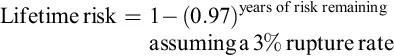
While the aforementioned studies have shown concordance in estimates of annual risk of rupture, variability among lesions and patient populations presents inherent challenges to generalizability and comprehensively predicting risk of rupture, as illustrated in various studies on the natural history of bAVMs (Pollock et al., 1996; Stapf et al., 2001; Stefani et al., 2002; Fullerton et al., 2005; Kim et al., 2010; Laakso et al., 2010; Gross and Du, 2013).
Classification systems
Multiple scales have been formulated to predict the morbidity and mortality associated with AVMs and the associated risk of intervention. Spetzler and Martin (1986), in their seminal paper on bAVMs, proposed a grading system based on size, venous drainage, and eloquence of brain adjacent to the malformation. The five-point scale assigns a score as follows: for size, <3 cm=1 point, 3–6 cm=2 points, >6 cm=3 points; for location, eloquent brain=1 point, noneloquent=0 points; for venous drainage, deep venous drainage=1 point, not deep venous drainage=0 points. Applying this scheme, higher scores translate to higher Spetzler–Martin grade, which corresponds to greater risk of morbidity and mortality with microsurgical removal of the lesion. (See Figure 1.1 for an example of Spetzler–Martin grade 3 AVM on preoperative imaging, intraoperative photos, and intraoperative fluorescence angiography processing.)

While the Spetzler–Martin grade is the most-quoted benchmark/standard in determining outcome and management, other grading systems have been introduced as adjuncts. Lawton et al. proposed a supplementary and complementary system to the Spetzler–Martin grade in 2010 that takes into account age, hemorrhagic presentation, and diffuseness of lesion, and yields improved accuracy in predictability of neurologic outcome when combined with the Spetzler–Martin scale (Table 1.1). This supplementary scale in conjunction with Spetzler–Martin is a 10-point scale that takes the five-point Spetzler–Martin scheme and, in addition, accounts for the following: for age, <20 years=1 point, 20–40=2 points, >40=3 points; for hemorrhagic presentation, hemorrhage=0 points, no hemorrhage=1 point; for lesion nidus diffuseness, compact=0 points, diffuse=1 point. Lawton et al. (2010) have shown that application of their scale can help with preoperative risk prediction and extrapolation of outcome (Kim et al., 2014).
Table 1.1
Comparing the Spetzler...
Table of contents
- Cover image
- Title page
- Table of Contents
- Copyright
- Handbook of Clinical Neurology 3rd Series
- Foreword
- Preface
- Contributors
- Part I: Arteriovenous Malformations
- Part II: Cavernous Malformations
- Index
Frequently asked questions
Yes, you can cancel anytime from the Subscription tab in your account settings on the Perlego website. Your subscription will stay active until the end of your current billing period. Learn how to cancel your subscription
No, books cannot be downloaded as external files, such as PDFs, for use outside of Perlego. However, you can download books within the Perlego app for offline reading on mobile or tablet. Learn how to download books offline
Perlego offers two plans: Essential and Complete
- Essential is ideal for learners and professionals who enjoy exploring a wide range of subjects. Access the Essential Library with 800,000+ trusted titles and best-sellers across business, personal growth, and the humanities. Includes unlimited reading time and Standard Read Aloud voice.
- Complete: Perfect for advanced learners and researchers needing full, unrestricted access. Unlock 1.4M+ books across hundreds of subjects, including academic and specialized titles. The Complete Plan also includes advanced features like Premium Read Aloud and Research Assistant.
We are an online textbook subscription service, where you can get access to an entire online library for less than the price of a single book per month. With over 1 million books across 990+ topics, we’ve got you covered! Learn about our mission
Look out for the read-aloud symbol on your next book to see if you can listen to it. The read-aloud tool reads text aloud for you, highlighting the text as it is being read. You can pause it, speed it up and slow it down. Learn more about Read Aloud
Yes! You can use the Perlego app on both iOS and Android devices to read anytime, anywhere — even offline. Perfect for commutes or when you’re on the go.
Please note we cannot support devices running on iOS 13 and Android 7 or earlier. Learn more about using the app
Please note we cannot support devices running on iOS 13 and Android 7 or earlier. Learn more about using the app
Yes, you can access Arteriovenous and Cavernous Malformations by in PDF and/or ePUB format, as well as other popular books in Medicine & Neurology. We have over one million books available in our catalogue for you to explore.