- 366 pages
- English
- ePUB (mobile friendly)
- Available on iOS & Android
eBook - ePub
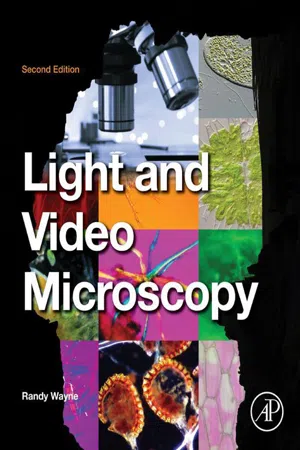
Light and Video Microscopy
About this book
The purpose of this book is to provide the most comprehensive, easy-to-use, and informative guide on light microscopy. Light and Video Microscopy will prepare the reader for the accurate interpretation of an image and understanding of the living cell. With the presentation of geometrical optics, it will assist the reader in understanding image formation and light movement within the microscope. It also provides an explanation of the basic modes of light microscopy and the components of modern electronic imaging systems and guides the reader in determining the physicochemical information of living and developing cells, which influence interpretation.
- Brings together mathematics, physics, and biology to provide a broad and deep understanding of the light microscope
- Clearly develops all ideas from historical and logical foundations
- Laboratory exercises included to assist the reader with practical applications
- Microscope discussions include: bright field microscope, dark field microscope, oblique illumination, phase-contrast microscope, photomicrography, fluorescence microscope, polarization microscope, interference microscope, differential interference microscope, and modulation contrast microscope
Tools to learn more effectively

Saving Books

Keyword Search

Annotating Text

Listen to it instead
Information
Chapter 1
The Relation Between the Object and the Image
In this chapter I discuss the relationship between the object and the image from a philosophical, a psychological, and a physical perspective. I do so by telling the story of Plato’s Allegory of the Cave and by showing optical illusions. I ask: (1) Can we be fooled by our eyes? and (2) How do we know the real nature of the object we see in the microscope? We will be able to know the real nature of the object if we understand the physics of light, the interaction of light with matter, and the action of the optical system of the microscope. I use the concept that light travels in straight lines to describe shadow formation and image formation by the pinhole in a camera obscura. I also discuss theories of vision. I describe the differences between luminous and nonluminous objects, and explain where light comes from and how it can be measured.
Keywords
Light; Luminous; Optical Illusions; Plato’s Cave; Vision
And God said, “Let there be light,” and there was light. God saw that the light was good, and he separated the light from the darkness.
Gen. 1:3-4
Since we acquire a significant amount of reliable information regarding the real world through our eyes, we often say, “seeing is believing.” However, seeing involves a number of processes that take place in space and time as light travels from a real object to our eyes and then gets coded into electrical signals that travel through the optic nerve to the brain. In the brain, neural signals are processed by the visual cortex, and ultimately the brain projects its interpretation of the real object as a virtual image seen by the mind’s eye. To ensure that “seeing is not deceiving” requires an understanding of light, optics, the interaction of light with matter, and how the brain functions to create and interpret the relationship between a real object and its image. According to Samuel Tolansky (1964), “There is often a failure in co-ordination between what we see and what we evaluate … .surprisingly enough, we shall find that most serious errors can creep even into scientific observations entirely because we are tricked by optical illusions into making quite faulty judgments.” Simon Henry Gage (1941), author of seventeen editions of the classic textbook, The Microscope, reminds us that the “image, whether it is made with or without the aid of the microscope, must always depend upon the character and training of the seeing and appreciating brain behind the eye.”
The light microscope, one of the most elegant instruments ever invented, is a device that permits us to study the interaction of light with matter at a resolution much greater than that of the unaided eye (Dobell, 1932; Wilson, 1995; Ruestow, 2004; Schickore, 2007; Ratcliff, 2009). Due to the constancy of the interaction of light with matter, we can peer into the would-be invisible world to discover the hidden properties of objects in that world (Appendix I). We can make transparent and invisible cells visible with a dark-field, phase-contrast, or differential interference microscope. We can use a polarizing microscope to reveal the orientation of macromolecules in a cell, and we can use it to determine the entropy and enthalpy of the polymerization process. We can use an interference microscope to weigh objects and to ascertain the mass of the cell’s nucleus. We can use a fluorescence microscope to localize proteins in the cytoplasm, genes on a chromosome, and the free Ca2+ concentration and pH of the surrounding milieu. We can use a centrifuge microscope or a microscope with laser tweezers to measure the forces involved in cellular motility or to determine the elasticity and viscosity of the cytoplasm. We can use a laser Doppler microscope, which takes advantage of the Doppler effect produced by moving objects, to characterize the velocities of organelles moving through the cytoplasm. We can also use a variety of laser microscopes to visualize single molecules.
I wrote this book so that you can make the most of the light microscope when it comes to faithfully creating and correctly interpreting images. To this end, the goals of this book are to:
• Describe the nature of light.
• Describe the relationship between an object and its image.
• Describe how light interacts with matter to yield information about the structure, composition, and local environment of biological and other specimens.
• Describe how optical systems work so that you will be able to interpret the images obtained at high resolution and magnification.
• Give you the necessary procedures and tricks so that you can gain practical experience with the light microscope and become an excellent microscopist.
Luminous and Nonluminous Objects
All objects, which are perceived by our sense of sight, can be divided into two broad classes. One class of objects, known as luminous bodies, includes “hot” or incandescent sources such as the sun, the stars, torches, oil lamps, candles, coal and natural gas lamps, kerosene lamps, and electric light bulbs, and “cold” sources such as fireflies and glow worms that produce “living light” (Brewster, 1830; Hunt, 1850; Harvey, 1920, 1940). These luminous objects are visible to our eyes. The second class of objects is nonluminous. However, they can be made visible to our eyes when they are in the presence of a luminous body. Thus the sun makes the moon, Earth, and other planets visible to us, and a light bulb makes all the objects in a room or on a microscope slide visible to us. The nonluminous bodies become visible by scattering the light that comes from luminous bodies. A luminous or nonluminous body is visible to us only if there are sufficient differences in brightness or color between it and its surroundings. The difference in brightness or color between points in the image formed of an object on our retina is known as contrast.
Object and Image
Each object is composed of many small and finite points composed of atoms or molecules. Ultimately, the image of each object is a point-by-point representation of that object upon our retina. Each point in the image should be a faithful representation of the brightness and color of the conjugate point in the object. Two points on different planes are conjugate if they represent identical spatial locations on the two planes. The object we see may itself be an intermediate image of a real object. The intermediate image of a real object observed with a microscope, telescope, or by looking at a photograph, movie, television screen, or computer monitor should also be a faithful point-by-point representation of the brightness and color of each conjugate point of the real object. While we only see brightness and color, the mind interprets the relative brightness and colors of the points of light on the retina and makes a judgment as to the size, shape, location, and position of the real object in its environment.
What we see, however, is not a perfect representation of the physical world. First, our eyes are not perfect, and our vision is limited by physical, genetic, and nutritional factors (Wald, 1967; Helmholtz, 2005). For example, we cannot see clearly things that are too far or too close, too dark or too bright, or things that emit radiation outside the visible range of wavelengths. Second, our vision is affected by physiological and psychological factors, and we can be easily fooled by our sense of sight (Goethe, 1840; Sully, 1881; Gregory, 1973; Békésy, 1967; Wade, 1998; Russ, 2004). Third, as Goethe learned when he studied the colors of the Italian landscape as they transformed from vibrant to muted and back again as the weather changed (Heisenberg, 1979), or as humankind learned upon the introduction of artificial illumination (Wickenden, 1910; Steinmetz, 1918; Otter, 2008), we must remember to take the source of illumination as well as the environment surrounding the object into consideration.
The architects of ancient Greece knew that the optical illusions that occur under certain circumstances, if not taken into consideration, would diminish the beauty of great buildings such as the Parthenon, which was built in honor of the virgin (parthenos) Athena (Penrose, 1851; Fletcher and Fletcher, 1905; Prokkola, 2011). For example, stylobates, or long horizontal foundations for the classical columns, and architraves, the horizontal beams above doorways, would appear to sag in the middle if they were made perfectly straight. Consequently, the architects used horizontal beams with convex tops to compensate for the optical illusion—the result being a perfectly square-looking structure. The columns of the Parthenon are famous, but they are not identical. The columns that are viewed against the bright Greek sky were made thicker than the columns backed by the inner temple or cella wall, since identical columns viewed against a bright background appear thinner than those viewed against a dark background. By compensating for the optical illusion, the columns appear identical and magnificent.
The sculptors of ancient Greece also knew about optical illusions, as evidenced by an apocryphal legend concerning two sculptors, Phidias, the teacher, and his student, Alkamenes (Anon, 1851). They were contenders in a contest to produce a sculpture of Athena that would stand upon a pedestal. Alkamenes sculpted a beautiful and well-proportioned figure of Athena, while Phidias, using his knowledge of geometry and optics, fashioned a grotesque and distorted figure. While the two sculptures were on the ground, the judges marveled at the one created by Alkamenes and laughed at the one created by Phidias. However, once the sculptures were put on top of the column, the perspective changed, and Phidias’s sculpture assumed great beauty while Alkamenes sculpture looked distorted. Knowing that the angles subtended by e...
Table of contents
- Cover image
- Title page
- Table of Contents
- Copyright
- Dedication
- Preface to the Second Edition
- Preface to the First Edition
- Chapter 1. The Relation Between the Object and the Image
- Chapter 2. The Geometric Relationship Between Object and Image
- Chapter 3. The Dependence of Image Formation on the Nature of Light
- Chapter 4. Bright-Field Microscopy
- Chapter 5. Photomicrography
- Chapter 6. Methods of Generating Contrast
- Chapter 7. Polarization Microscopy
- Chapter 8. Interference Microscopy
- Chapter 9. Differential Interference Contrast (DIC) Microscopy
- Chapter 10. Amplitude Modulation Contrast Microscopy
- Chapter 11. Fluorescence Microscopy
- Chapter 12. Various Types of Microscopes and Accessories
- Chapter 13. Video and Digital Microscopy
- Chapter 14. Image Processing and Analysis
- Chapter 15. Laboratory Exercises
- References
- Appendix I. Light Microscopy: A Retrospective
- Appendix II. A Final Exam
- Appendix III. A Microscopist’s Model of the Photon
- Index
- Color Plates
Frequently asked questions
Yes, you can cancel anytime from the Subscription tab in your account settings on the Perlego website. Your subscription will stay active until the end of your current billing period. Learn how to cancel your subscription
No, books cannot be downloaded as external files, such as PDFs, for use outside of Perlego. However, you can download books within the Perlego app for offline reading on mobile or tablet. Learn how to download books offline
Perlego offers two plans: Essential and Complete
- Essential is ideal for learners and professionals who enjoy exploring a wide range of subjects. Access the Essential Library with 800,000+ trusted titles and best-sellers across business, personal growth, and the humanities. Includes unlimited reading time and Standard Read Aloud voice.
- Complete: Perfect for advanced learners and researchers needing full, unrestricted access. Unlock 1.4M+ books across hundreds of subjects, including academic and specialized titles. The Complete Plan also includes advanced features like Premium Read Aloud and Research Assistant.
We are an online textbook subscription service, where you can get access to an entire online library for less than the price of a single book per month. With over 1 million books across 990+ topics, we’ve got you covered! Learn about our mission
Look out for the read-aloud symbol on your next book to see if you can listen to it. The read-aloud tool reads text aloud for you, highlighting the text as it is being read. You can pause it, speed it up and slow it down. Learn more about Read Aloud
Yes! You can use the Perlego app on both iOS and Android devices to read anytime, anywhere — even offline. Perfect for commutes or when you’re on the go.
Please note we cannot support devices running on iOS 13 and Android 7 or earlier. Learn more about using the app
Please note we cannot support devices running on iOS 13 and Android 7 or earlier. Learn more about using the app
Yes, you can access Light and Video Microscopy by Randy O. Wayne in PDF and/or ePUB format, as well as other popular books in Scienze biologiche & Biologia. We have over one million books available in our catalogue for you to explore.