- 418 pages
- English
- ePUB (mobile friendly)
- Available on iOS & Android
eBook - ePub
Guide to Research Techniques in Neuroscience
About this book
Neuroscience is, by definition, a multidisciplinary field: some scientists study genes and proteins at the molecular level while others study neural circuitry using electrophysiology and high-resolution optics. A single topic can be studied using techniques from genetics, imaging, biochemistry, or electrophysiology. Therefore, it can be daunting for young scientists or anyone new to neuroscience to learn how to read the primary literature and develop their own experiments.
This volume addresses that gap, gathering multidisciplinary knowledge and providing tools for understanding the neuroscience techniques that are essential to the field, and allowing the reader to design experiments in a variety of neuroscience disciplines.
- Written to provide a "hands-on" approach for graduate students, postdocs, or anyone new to the neurosciences
- Techniques within one field are compared, allowing readers to select the best techniques for their own work
- Includes key articles, books, and protocols for additional detailed study
- Data analysis boxes in each chapter help with data interpretation and offer guidelines on how best to represent results
- Walk-through boxes guide readers step-by-step through experiments
Information
Chapter 1
Whole-Brain Imaging
Abstract
The purpose of this chapter is to explain the ostensible magic of whole-brain imaging technology and provide insight into how experiments are designed and interpreted. This chapter will give you the information needed so you may compare the relative strengths and limitations of different structural and functional brain imaging techniques, explain the physical and physiological basis of magnetic resonance imaging/functional magnetic resonance imaging technology, and describe the design of a functional brain imaging experiment: formulating a hypothesis, choosing task paradigms, performing the experiment, acquiring and analyzing data, and constructing figures.
Keywords
Cerebral angiography; Computerized tomography (CT); Diffusion MR imaging; Electroencephalography (EEG); Functional magnetic resonance imaging (fMRI); Magnetic resonance imaging (MRI); Magnetoencephalography (MEG); Optical imaging; Positron emission tomography (PET); Single-proton emission computerized tomography (SPECT); Transcranial magnetic stimulation (TMS); Ultrasonic neuromodulation (USNM)After Reading This Chapter, You Should be Able to:
• Compare the relative strengths and limitations of different structural and functional brain imaging techniques
• Explain the physical and physiological basis of (MRI/fMRI) technology
• Describe the design of a functional brain imaging experiment: formulating a hypothesis, choosing task paradigms, performing the experiment, acquiring and analyzing data, and constructing figures
Techniques covered:
• Structural techniques: cerebral angiography, computerized tomography (CT), MRI, and diffusion MR imaging
• Functional techniques: fMRI, positron emission tomography (PET), single-proton emission computerized tomography (SPECT), electroencephalography (EEG), magnetoencephalography (MEG), and optical imaging
• Techniques used to investigate the necessity and sufficiency of a specific brain region for a cognitive function: transcranial magnetic stimulation (TMS), ultrasonic neuromodulation (USNM), and case studies
Modern brain imaging technology can seem like magic. The ability to produce detailed images of the human brain without physically penetrating the skull is a technological marvel that has saved thousands of lives and allowed scientists to study the structure of the brain throughout development, disease, and aging. Furthermore, the ability to image neural activity in the brain during cognition has provided scientists the opportunity to correlate activity in distinct brain regions with specific mental operations, a truly remarkable achievement. Colorful figures depicting activity in the human brain dazzle scientists and nonscientists alike.
Of course, brain imaging technology is not magic. The technology that produces detailed images of the brain depends on complex physics, expensive equipment, and skilled technicians. As with all scientific experiments, brain imaging studies must be well designed, the data accurately analyzed, and the results carefully interpreted. The purpose of this chapter is to explain the ostensible magic of whole-brain imaging technology and provide insight into how experiments are designed and interpreted.
Whole-brain imaging technology can essentially be divided into two categories: structural and functional. Structural techniques produce images of the anatomical architecture of the brain, whereas functional techniques produce images of the physiological processes that underscore neural activity. This chapter will survey both classifications of techniques and describe how they can be used in modern neuroscience research. We focus on MRI and fMRI technology in humans due to the widespread use of these techniques in the neuroscience literature. After reviewing these techniques, we will survey the essential components of a functional imaging experiment: forming hypotheses, choosing appropriate task paradigms, performing experiments, acquiring and analyzing data, and producing figures for publication.
Structural Brain Imaging Techniques
Structural brain imaging techniques are used to resolve the anatomy of the brain in a living subject without physically penetrating the skull. These techniques can be used in combination with functional brain imaging techniques to correlate neural activity in specific anatomical regions with behavioral or cognitive functions. Structural techniques can also be used to measure anatomical changes that occur over time, such as a decrease in brain mass that occurs with aging or the progression of disease. Most often, these techniques are used in clinical neuroscience and neurology to diagnose diseases such as tumors and vascular disorders.
Brain imaging technologies take advantage of the different biochemical compositions of brain regions and use these differences to form the basis of an image (Figure 1.1). Neural cell bodies contain many biomolecules, including proteins and carbohydrates. Axons and fiber tracts are relatively fatty due to the insulation provided by myelin. Cerebrospinal fluid (CSF) in the ventricles and surrounding the brain is essentially a saline solution. The microanatomy and composition of individual neural structures cause distinct regions of the brain to appear different when examined with the naked eye. For example, when examining slices of a brain, tissue mostly composed of cell bodies appears gray compared with other areas and therefore is referred to as “gray matter.” Brain tissue mostly composed of axons and fiber tracts appears white and therefore is referred to as “white matter.” Often, the most informative structural images of the brain show the contrast between gray and white matter. Thus, the ultimate goal of structural imaging technologies is to differentiate between proteins and carbohydrates (cell bodies), fat (axon tracts), and salt water (CSF), as this contrast reveals the most information about brain architecture.
Until the early 1970s, there was no technology that could differentiate between these substances within the brain. Conventional X-ray technology is essentially useless for this purpose. During an X-ray procedure, an X-ray beam is passed through an object and then onto a photographic plate (Figure 1.2A). Each of the molecules through which the beam passes absorbs some of the radiation, so only the unabsorbed portions of the beam reach the photographic plate. X-ray photography is, therefore, only effective in characterizing internal structures that differ substantially from their surroundings in the degree to which they absorb X-rays, such as bone in flesh (Figure 1.2B). By the time an X-ray beam passes through the relatively soft consistency of the brain (not to mention the relatively hard consistency of the skull!), little information about individual brain structures can be discerned (Figure 1.2C). Therefore, in the 1960s and 1970s, there was strong motivation to discover better ways of imaging the brain. The techniques described in the following sections represent 30–40 years worth of innovation in technology ultimately designed to show contrast within the soft tissue of the brain.
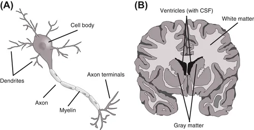
Figure 1.1 The composition of the brain.
(A) A microscopic view of a neuron. Each neuron is composed of a cell body, dendrites, and an axonal process. The cell bodies and dendrites are rich in proteins and carbohydrates. Axons are surrounded by a myelin insulation made of fats. (B) A macroscopic view of the brain. Gray matter is rich in cell bodies and, therefore, in proteins and carbohydrates. White matter is composed of axon tracts and is, therefore, rich in fatty myelin. Cerebrospinal fluid (CSF) in the ventricles is a saline solution. To produce an image, brain imaging technologies must differentiate among proteins/carbohydrates, fats, and saline.
Cerebral Angiography
A cerebral angiogram is an enhanced X-ray that uses dyes to make up for the relatively poor soft-tissue contrast of conventional X-rays. A radio-opaque dye that absorbs X-rays better than surrounding tissue is injected into an artery that delivers blood to the brain. This substance heightens the contrast between the cerebral circulatory system and surrounding brain tissue during an X-ray (Figure 1.3A). Thus, the most prominent aspect of the central nervous system imaged in a cerebral angiogram is the brain vasculature. Angiograms can show vascular damage and indicate the presence of a tumor or aneurysm, an abnormal ballooning of a portion of an artery due to weakening of a blood vessel wall (Figure 1.3B).
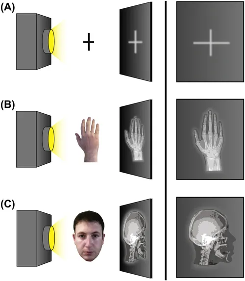
Figure 1.2 Standard X-ray technology alone cannot produce detailed images of the brain.
(A) During an X-ray procedure, an X-ray beam is passed through an object and onto a photographic plate. Only the unabsorbed portions of the beam reach the plate, creating an image. (B) The contrast between the soft consistency of skin and muscle compared with the hard consistency of bone is sufficient to form an X-ray image. However, (C) the contrast between the soft consistency of different tissues within the nervous system is insufficient to form an X-ray image of the brain.
Computerized Tomography
Another method that improves upon conventional X-ray technology to image the brain and body is computerized tomography (the “CT scan”—sometimes also called computerized axial tomo...
Table of contents
- Cover image
- Title page
- Table of Contents
- Copyright
- Foreword to the Second Edition
- Foreword to the First Edition
- Preface to the Second Edition
- Preface to the First Edition
- Introduction
- Chapter 1. Whole-Brain Imaging
- Chapter 2. Animal Behavior
- Chapter 3. Stereotaxic Surgeries and In Vivo Techniques
- Chapter 4. Electrophysiology
- Chapter 5. Microscopy
- Chapter 6. Visualizing Neural Structure
- Chapter 7. Visualizing Neural Function
- Chapter 8. Manipulating Neural Activity
- Chapter 9. Identifying Genes and Proteins of Interest
- Chapter 10. Molecular Cloning and Recombinant DNA Technology
- Chapter 11. Gene Delivery Strategies
- Chapter 12. Making and Using Transgenic Organisms
- Chapter 13. Manipulating Endogenous Genes
- Chapter 14. Cell Culture Techniques
- Chapter 15. Biochemical Assays and Intracellular Signaling
- Glossary
- Index
Frequently asked questions
Yes, you can cancel anytime from the Subscription tab in your account settings on the Perlego website. Your subscription will stay active until the end of your current billing period. Learn how to cancel your subscription
No, books cannot be downloaded as external files, such as PDFs, for use outside of Perlego. However, you can download books within the Perlego app for offline reading on mobile or tablet. Learn how to download books offline
Perlego offers two plans: Essential and Complete
- Essential is ideal for learners and professionals who enjoy exploring a wide range of subjects. Access the Essential Library with 800,000+ trusted titles and best-sellers across business, personal growth, and the humanities. Includes unlimited reading time and Standard Read Aloud voice.
- Complete: Perfect for advanced learners and researchers needing full, unrestricted access. Unlock 1.4M+ books across hundreds of subjects, including academic and specialized titles. The Complete Plan also includes advanced features like Premium Read Aloud and Research Assistant.
We are an online textbook subscription service, where you can get access to an entire online library for less than the price of a single book per month. With over 1 million books across 990+ topics, we’ve got you covered! Learn about our mission
Look out for the read-aloud symbol on your next book to see if you can listen to it. The read-aloud tool reads text aloud for you, highlighting the text as it is being read. You can pause it, speed it up and slow it down. Learn more about Read Aloud
Yes! You can use the Perlego app on both iOS and Android devices to read anytime, anywhere — even offline. Perfect for commutes or when you’re on the go.
Please note we cannot support devices running on iOS 13 and Android 7 or earlier. Learn more about using the app
Please note we cannot support devices running on iOS 13 and Android 7 or earlier. Learn more about using the app
Yes, you can access Guide to Research Techniques in Neuroscience by Matt Carter,Jennifer C. Shieh in PDF and/or ePUB format, as well as other popular books in Medicine & Neurology. We have over one million books available in our catalogue for you to explore.