- English
- ePUB (mobile friendly)
- Available on iOS & Android
Interleukin-1 in the Brain
About this book
Interest in interleukin-1 (IL-1) has increased dramatically over the last decade, but has been largely restricted to immunologists, cell biologists and those studying inflammation and cancer. However, it has recently been recognized that the brain directly controls or modulates many aspects of immune function, while molecules classically associated with the immune system, such as interleukin-1, are synthesised within the brain and act directly on the central nervous system to modify local and systemic functions. Thus, this topic is relatively new to neurobiologists, and this book is the first comprehensive description of current knowledge on interleukin-1 in the brain, including its location, synthesis and receptors, actions on behaviour, fever, metabolism, neuroendocrine function, electrical activity of the brain, nerve growth factor, and relationship to clinical indications. The book is organised into three sections. The first reviews the data available on the neural localisation of IL-1 and the nature of its central receptors. The main part of the book examines the different neural effects of IL-1 and the mechanisms which are involved in these actions, comparing IL-1 where possible to other inflammatory cytokines which also have neurotrophic effects. The final section evaluates the possible role of IL-1 in neural plasticity and neuronal degeneration.
Tools to learn more effectively

Saving Books

Keyword Search

Annotating Text

Listen to it instead
Information
Location of interleukin-1 in the nervous system
Publisher Summary
1.1 IL-1 in the peripheral nervous system
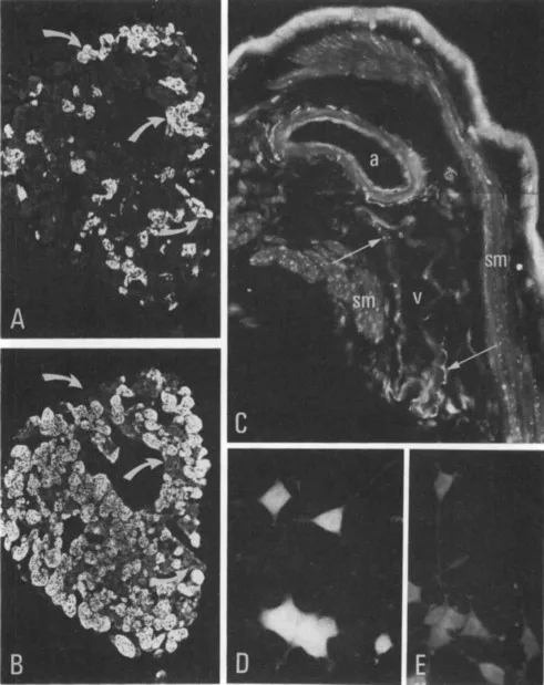
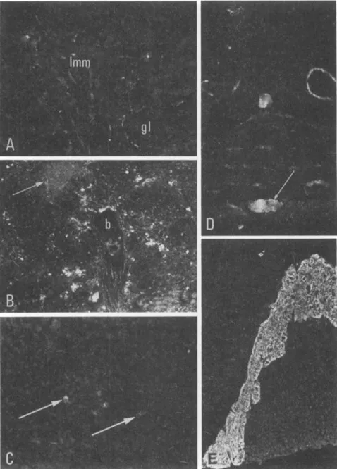
Table of contents
- Cover image
- Title page
- Table of Contents
- Pergamon studies in neuroscience No 5
- Copyright
- FOREWORD
- Chapter 1: Location of interleukin-1 in the nervous system
- Chapter 2: Brain interleukin-1 receptors: mapping, characterization and modulation
- Chapter 3: Interleukin-1β activation of the central nervous system
- Chapter 4: Electrophysiological studies of the effects of interleukin-1 and α-interferon on the EEG and pituitary-adrenocortical activity
- Chapter 5: The immune-hypothalamo-pituitary adrenal axis: Its role in immunoregulation and tolerance to self-antigens
- Chapter 6: The pyrogenic action of cytokines
- Chapter 7: Metabolic responses to interleukin-1
- Chapter 8: Behavioural effects of cytokines
- Chapter 9: Interleukin-1 involvement in the regulation of sleep
- Chapter 10: Regulation of the synthesis of nerve growth factor (NGF) by interleukin-1 (IL-1): facts and questions
- Chapter 11: Cytokines and neuronal degeneration
- INDEX
Frequently asked questions
- Essential is ideal for learners and professionals who enjoy exploring a wide range of subjects. Access the Essential Library with 800,000+ trusted titles and best-sellers across business, personal growth, and the humanities. Includes unlimited reading time and Standard Read Aloud voice.
- Complete: Perfect for advanced learners and researchers needing full, unrestricted access. Unlock 1.4M+ books across hundreds of subjects, including academic and specialized titles. The Complete Plan also includes advanced features like Premium Read Aloud and Research Assistant.
Please note we cannot support devices running on iOS 13 and Android 7 or earlier. Learn more about using the app