Microbial Glycobiology
eBook - ePub

Microbial Glycobiology

Structures, Relevance and Applications

  1. 1,036 pages
  2. English
  3. ePUB (mobile friendly)
  4. Available on iOS & Android
eBook - ePub

Microbial Glycobiology

Structures, Relevance and Applications

Book details
Book preview
Table of contents
Citations

About This Book

This book presents in an easy-to-read format a summary of the important central aspects of microbial glycobiology, i.e. the study of carbohydrates as related to the biology of microorganisms. Microbial glycobiology represents a multidisciplinary and emerging area with implications for a range of basic and applied research fields, as well as having industrial, medical and biotechnological implications.

  • Individual chapters provided by leading international scientists in the field yield insightful, concise and stimulating reviews
  • Provides researchers with an overview and synthesis of the latest research
  • Each chapter begins with a brief 200 word Summary/Abstract detailing the topic and focus of the chapter, as well as the concepts to be addressed
  • Allows researchers to see at a glance what each chapter will cover
  • Each chapter includes a Research Focus Box
  • Identifies important problems that still need to be solved and areas that require further investigation

Frequently asked questions

Simply head over to the account section in settings and click on ā€œCancel Subscriptionā€ - itā€™s as simple as that. After you cancel, your membership will stay active for the remainder of the time youā€™ve paid for. Learn more here.
At the moment all of our mobile-responsive ePub books are available to download via the app. Most of our PDFs are also available to download and we're working on making the final remaining ones downloadable now. Learn more here.
Both plans give you full access to the library and all of Perlegoā€™s features. The only differences are the price and subscription period: With the annual plan youā€™ll save around 30% compared to 12 months on the monthly plan.
We are an online textbook subscription service, where you can get access to an entire online library for less than the price of a single book per month. With over 1 million books across 1000+ topics, weā€™ve got you covered! Learn more here.
Look out for the read-aloud symbol on your next book to see if you can listen to it. The read-aloud tool reads text aloud for you, highlighting the text as it is being read. You can pause it, speed it up and slow it down. Learn more here.
Yes, you can access Microbial Glycobiology by Anthony P Moran, Otto Holst,Patrick Brennan,Mark von Itzstein in PDF and/or ePUB format, as well as other popular books in Biological Sciences & Biochemistry. We have over one million books available in our catalogue for you to explore.

Information

Year
2009
ISBN
9780080923246
Chapter 1. Overview of the glycosylated components of the bacterial cell envelope
Otto Holst, Anthony P. Moran and Patrick J. Brennan

Summary

Within this chapter, the various types of bacterial cell envelope and their carbohydrate-related molecules, as well as the glycoprotein S-layers that can be found in all bacteria except mycobacteria, are introduced. Whereas each of the Gram-positive, Gram-negative and mycobacterial cell envelopes possess a general and typical architecture, the archaeal cell envelope shows a broader variety of constructions and may be comprised of only a membrane. Apart from S-layers, carbohydrate-containing macromolecules like lipopolysaccharides, peptidoglycan, lipoteichoic acids, teichoic acids, capsule polysaccharides, lipoarabinomannan and others are briefly described. This chapter refers to other, more detailed, subsequent chapters that summarize the chemistry and biological function of the cell envelope macromolecules.
Keywords: Bacterial cell envelope; Lipopolysaccharide; Lipoteichoic acid; Teichoic acid; Lipoarabinomannan; Arabinogalactan; Peptidoglycan; Polysaccharide; Glycolipid; Glycoprotein

1. Introduction ā€“ The Bacterial Cell Envelope Encountering Environmental Challenges

Bacteria of various species populate most of the environments encountered on Earth. As a group, these microorganisms cope with very low, even down to āˆ’4Ā°C, temperatures (psycrophilic bacteria), medium range temperatures (mesophilic bacteria), warm to hot temperatures (thermophilic bactera) or very hot, up to 115Ā°C, temperatures (hyperthermophilic bacteria). Also, they exhibit an ability to survive and withstand an impressive range of pH, between pH 0.7 and 9 (acido- and alkaliphilic bacteria), to survive high pressures (deep sea, barophilic bacteria), or they may need or have to survive high or higher salt concentrations (halotolerant and halophilic bacteria) in their environments. Moreover, they may need to live and proliferate in eukaryotic hosts (symbiotic or pathogenic bacteria). In addition, bacterial species have developed the means to overcome longer periods of dryness and/or starvation or exposure to strong UV radiation. In order to survive in a particular niche, bacteria have developed a large variety of intracellular physiological adaptations; also, most possess an outer cellular barrier, the cell envelope, by which they communicate with and protect themselves from the environment they inhabit (Seltmann and Holst, 2001).
In nearly all genera of the domain Bacteria and in several of those of the domain archaeal, this layer is present which, by definition, consists of the cytoplasmic membrane (CM), the cell wall and, if present, outer layers such as capsules or sheaths. The outer barrier of other genera of the domain archaeal is formed by a particular membrane structure which contains ether-linked lipids that span the whole membrane and are highly resistant to temperature and pH (Seltmann and Holst, 2001).
When considering the broad variety of environments inhabited and encountered by bacteria, it is quite astonishing that only a few general architectural forms of cell envelope have evolved to cope with these varied conditions. The common architectural principle of the CM and cell wall is present in all bacterial envelopes in the four great variations of the envelope, i.e. Gram-negative bacterial, Gram-positive bacterial, mycobacterial and archaeal, and which may possess further adaptation even at the species level. Importantly, all cell envelopes possess a significant proportion of carbohydrate-containing constituents. This chapter serves to introduce the various types of cell envelope and their carbohydrate-related molecules and the glycoprotein S-layers that can be found in all bacteria except mycobacteria. As subsequent chapters will present more detailed reviews of the cell envelope-related macromolecules, their synthesis and applications, extensive literature will not be cited here, merely the relevant chapters will be indicated.

2. The Gram-Negative Cell Envelope

The Gram-negative bacterial world contains a broad variety of genera that may comprise only harmless species (e.g. phototrophic bacteria of genera like Rhodobacter or Rhodomicrobium) or both harmless and human- or animal-pathogenic species (e.g. Escherichia coli or Acinetobacter spp.) or plant-pathogenic species (e.g. Erwinia and Xanthomonas spp.). Independent of this, all Gram-negative bacteria possess a cell envelope of the same general architecture which is schematically depicted in Figure 1.1. The periplasmic ā€œspaceā€ is present on top of the CM and contains peptidoglycan (PG), also known as murein, as a major constituent. This macromolecular sacculus is composed of sugar chains built from alternating 2-acetamido-2-deoxy-d-glucopyranose (GlcpNAc) and 2-acetamido-3-O-[(R)-1-carboxyethyl]-2-deoxy-d-glucopyranose, also termed N-acetylmuramic acid (MurNAc), residues which carry, and can be cross-linked by, smaller peptides ā€“ the amino acid composition of which varies in different species. The PG sacculus represents a rigid layer that determines the form of the bacterial cell and is important for osmotic stability and acts as a protective barrier. However, the PG sacculus is not a completely closed wall that inhibits transport of small molecules (e.g. nutrients) or their excretion. Although very stable, the PG matrix represents a mesh with holes large enough to guarantee the flow of molecules, including those that have to be transported to the outer membrane (OM). The three-dimensional architecture of PG has been an issue of discussion for a long time. The first model, which many scientists consider represents the correct one, is built from sugar chains that run in a parallel direction to the CM and which are interlinked by short peptide stems that are oriented perpendicular to this membrane. About ten years ago, an alternative, called the scaffold model, was proposed with perpendicular sugar chains connected by peptides that run parallel to the CM. It should be noted that there is no clear proof for either model to date. Importantly, the Gram-negative PG is a rather thin construction making its examination difficult. A detailed review of PG is given in Chapter 2 and of its biosynthesis in Chapter 16.
B9780123745460000018/gr1.webp is missing
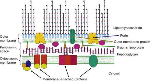
Figure 1.1
A schematic model of the Gram-negative cell envelope as present in E. coli. The cell envelope comprises the cytoplasmic membrane, which is a symmetric membrane consisting mainly of phospholipids in both leaflets, the periplasmic space and the outer membrane. Embedded in the cytoplasmic membrane, i.e. membrane-attached at the cytosolic leaflet or integrated into the membrane, are a number of proteins including those important for lipopolysaccharide or capsular polysaccharide biosynthesis and transport to the periplasmic space. The thin peptidoglycan layer represents the major constituent of the periplasmic space, but which also contains proteins including transport-proteins (indicated by the ovoid shapes in the periplasm). In E. coli, Braunā€™s lipoprotein is present which is covalently bound to the peptidoglycan and anchored by its lipid moiety in the inner leaflet of the outer membrane. In contrast to the cytoplasmic membrane, the outer membrane represents an asymmetric membrane, i.e. comprising phospholipids in the inner and lipopolysaccharides in the outer leaflet. It contains a number of outer membrane proteins, including the porins that are water-filled channels important for the import of small molecules, like sugars or ions. Not shown are capsular polysaccharides and the enterobacterial common antigen which may also be anchored in the outer leaflet by a lipid structure. In addition, S-layer glycoproteins may be present in certain Gram-negative bacterial species (not shown).
Apart from PG, the periplasmic space contains various smaller molecules like mono- and oligosaccharides, amino acids and peptides, as well as the biosynthetic precursors and degradation products of PG. The concentration of all these substances is rather high, thus the periplasmic space represents a highly viscous solution that can be considered a gel-like matrix.
Outside the periplasmic space is located a second membrane, the OM, which had been considered unique to Gram-negative bacteria for a long time. Nevertheless, the presence of an analogous structure in mycobacteria has been postulated for quite some time and whose presence has finally been verified recently (see below) (Hoffmann et al., 2008). Both leaflets of the OM represent lipid bilayers that are organized asymmetrically, i.e. the inner leaflet is composed of different molecules compared to the outer one. In the case of the OM of Gram-negative bacteria, the inner leaflet is comprised of phospholipids, whereas the outer leaflet is mainly constructed from lipopolysaccharides (LPSs) (Holst and MĆ¼ller-Loennies, 2007). In some cases, polysaccharide capsules are present that may be anchored by a lipid into the OM and this is also true for the enterobacterial common antigen (ECA) of enterobacteria. In addition, the OM contains various proteins (outer membrane proteins, OMPs), constituting up to 50% of the membrane and which often interact with LPS molecules, thereby yielding particular lipidā€“protein structural units. Several of the OMPs are channel-formers and are involved in diffusion of ions (e.g. diffusion of phosphate through PhoE channels in E. coli) and transport of mono- and small oligosaccharides (e.g. specific-channel forming proteins, like LamB that is involved in transport of maltose and maltodextrins) and, thus, they play important roles in the uptake of nutrients. Other OMPs represent structural proteins like OmpA and related proteins in various enterobacterial species and Braunā€™s lipoprotein, also known as PG-associated lipoprotein, in E. coli and related species. The latter is covalently linked to PG and has its lipid moiety embedded in the inner leaflet of the OM, thus interconnecting PG and OM and providing structural stability.
Molecules of LPS are of high relevance not only for Gram-negative bacteria but also for an infected, eukaryotic host. In bacteria, these molecules are part of the protective barrier shielding the microbes from dangerous environmental compounds, like bile salts in the gut or antibiotics in sensu latu. In contrast, domains within LPS, specifically the core region and the O-specific polysaccharide, can also act as receptors for bacteriophages, thereby contributing indirectly to the destruction of the bacterial cell in such cases. In pathogenic bacteria that infect either humans or animals or plants, LPSs represent very important virulence factors. Moreover, this family of molecules is also called the endotoxins of Gram-negative bacteria; however, their toxicity is highly dependent on their structural properties, and it should be noted that not all LPSs are toxic molecules, not even those from pathogens. Importantly, in many chronically infecting bacterial species (e.g. Helicobacter pylori, Porphyromonas gingivalis, etc.), LPSs are of low toxic and immunological act...

Table of contents

  1. Cover image
  2. Table of Contents
  3. Copyright
  4. Dedication
  5. List of Contributors
  6. Preface
  7. Chapter 1. Overview of the glycosylated components of the bacterial cell envelope
  8. Chapter 2. Bacterial cell envelope peptidoglycan
  9. Chapter 3. Core region and lipid A components of lipopolysaccharides
  10. Chapter 4. O-Specific polysaccharides of Gram-negative bacteria
  11. Chapter 5. Teichoic acids, lipoteichoic acids and related cell wall glycopolymers of Gram-positive bacteria
  12. Chapter 6. Bacterial capsular polysaccharides and exopolysaccharides
  13. Chapter 7. Bacterial surface layer glycoproteins and ā€œnon-classicalā€ secondary cell wall polymers
  14. Chapter 8. Glycosylation of bacterial and archaeal flagellins
  15. Chapter 9. Glycosylated components of the mycobacterial cell wall
  16. Chapter 10. Glycoconjugate structure and function in fungal cell walls
  17. Chapter 11. Cytoplasmic carbohydrate molecules
  18. Chapter 12. Glycosylated compounds of parasitic protozoa
  19. Chapter 13. Analytical approaches towards the structural characterization of microbial wall glycopolymers
  20. Chapter 14. Single-molecule characterization of microbial polysaccharides
  21. Chapter 15. Viral surface glycoproteins in carbohydrate recognition
  22. Chapter 16. Biosynthesis of bacterial peptidoglycan
  23. Chapter 17. Biosynthesis and membrane assembly of lipid A
  24. Chapter 18. Biosynthesis of O-antigen chains and assembly
  25. Chapter 19. Biosynthesis of cell wall teichoic acid polymers
  26. Chapter 20. Biosynthesis and assembly of capsular polysaccharides
  27. Chapter 21. Biosynthesis of the mycobacterial cell envelope components
  28. Chapter 22. Biosynthesis of fungal and yeast glycans
  29. Chapter 23. Chemical synthesis of bacterial lipid A
  30. Chapter 24. Chemical synthesis of the core oligosaccharide of bacterial lipopolysaccharide
  31. Chapter 25. Chemical synthesis of lipoteichoic acid and derivatives
  32. Chapter 26. Chemical synthesis of parasitic glycoconjugates and phosphoglycans
  33. Chapter 27. Bacterial lectin-like interactions in cell recognition and adhesion
  34. Chapter 28. Lectin-like interactions in virusā€“cell recognition
  35. Chapter 29. Sialic acid-specific microbial lectins
  36. Chapter 30. Bacterial toxins and their carbohydrate receptors at the hostā€“pathogen interface
  37. Chapter 31. Toll-like receptor recognition of lipoglycans, glycolipids and lipopeptides
  38. Chapter 32. NOD receptor recognition of peptidoglycan
  39. Chapter 33. Microbial interaction with mucus and mucins
  40. Chapter 34. Mannoseā€“fucose recognition by DC-SIGN
  41. Chapter 35. Host surfactant proteins in microbial recognition
  42. Chapter 36. T-Cell recognition of microbial lipoglycans and glycolipids
  43. Chapter 37. Extracellular polymeric substances in microbial biofilms
  44. Chapter 38. Physicochemical properties of microbial glycopolymers
  45. Chapter 39. Microbial biofilm-related polysaccharides in biofouling and corrosion
  46. Chapter 40. Microbial glycosylated components in plant disease
  47. Chapter 41. Antigenic variation of microbial surface glycosylated molecules
  48. Chapter 42. Phase variation of bacterial surface glycosylated molecules in immune evasion
  49. Chapter 43. Molecular mimicry of host glycosylated structures by bacteria
  50. Chapter 44. Role of microbial glycosylation in host cell invasion
  51. Chapter 45. Exopolysaccharides produced by lactic acid bacteria in food and probiotic applications
  52. Chapter 46. Industrial exploitation by genetic engineering of bacterial glycosylation systems
  53. Chapter 47. Glycomimetics as inhibitors in anti-infection therapy
  54. Chapter 48. Bacterial polysaccharide vaccines
  55. Chapter 49. Immunomodulation by zwitterionic polysaccharides
  56. Chapter 50. Future potential of glycomics in microbiology and infectious diseases
  57. Index