- 574 pages
- English
- ePUB (mobile friendly)
- Available on iOS & Android
eBook - ePub
Physiology and Biochemistry
About this book
The Structure and Function of Muscle, Second Edition, Volume III: Physiology and Biochemistry presents the physiology and biochemistry of muscle. This book discusses the various aspects of the structure of muscles and explores some aspects of muscle disease.
Organized into 10 chapters, this edition begins with an overview of the transverse tubular system or T system of striated muscle. This text then examines the properties and function of membranes through electron microscopy. Other chapters consider in more detail from a biophysical viewpoint certain aspects of the series of events surrounding muscle contraction. This book discusses as well the significance of the central circulation and the amount of oxygen that can be delivered by the cardiovascular system. The final chapter deals with the heat output and chemical breakdown during an isometric twitch.
This book is a valuable resource for scientists, neurobiologists, biologists, biochemists, physiologists, histologists, cytologists, and research workers.
Tools to learn more effectively

Saving Books

Keyword Search

Annotating Text

Listen to it instead
Information
1
ELECTRICAL PROPERTIES OF THE TRANSVERSE TUBULAR SYSTEM
LEE D. PEACHEY and RICHARD H. ADRIAN
Publisher Summary
This chapter discusses the electrical properties of the transverse tubular system. The electrical properties of nerve and muscle are analyzed in terms of the ionic and capacity currents flowing across the cell surface membrane. Morphological evidence suggests that the tubular system can be represented in frog twitch striated muscle fibers as a set of regular two-dimensional tubular networks in which the tubular diameter is smaller than the mesh and the mesh is smaller than the fiber diameter. Access resistance occurs because of constriction of the tubular lumen at the fiber surface or tortuosity of the tubular path near the surface. Either an increased tubular length or a reduced luminal caliber over a significant length increases the radial resistance near the fiber surface. The chapter also presents a linear and a nonlinear method for obtaining the spatial distribution of potential in the tubular system.
I. Introduction
II. Mathematical Analysis
A. Cable Analysis as a Network
B. Introduction of an Access Resistance
C. Tubular Length Constant and Resistance
D. Numerical Method of Analysis
III. Tubular Capacity—Lumped or Distributed?
IV. Excitation–Contraction Coupling
References
I Introduction
The transverse tubular system or T system of striated muscle was discovered by electron microscopy in the 1950s. The most important paper of that period (Porter and Palade, 1957) described the structure we now call the T system as rows of vesicles in the space between the terminal cisternae of the sarcoplasmic reticulum (SR) and termed it the “intermediary vesicles.” This analysis, in effect, included the T system as part of the SR. Since that time, through further comparative studies and the use of improved preparation methods, electron microscopists have demonstrated convincingly that the T system is a branched network of tubules derived from and remaining attached to the surface plasma membrane of the muscle fiber. This has fostered a general feeling that the T system should not be considered as a part of the SR, but as part of the surface membrane complex of the cell. To be sure, it is found deep in the fiber, and it associates closely and in a specific way with the intracellular SR, but in an important respect it is part of the fiber surface membrane.
Of greatest interest to us are the function and physiological properties of the T system. Several recent physiological and morphological studies have been directed to this problem, and work in this area continues actively at present. Our aim in this chapter will be to review the present state of our knowledge, and to indicate some of the uncertainties and possible future directions for advance. We also hope to resolve some uncertainties and apparent discrepancies in earlier papers by presenting an analysis of the T system as a cable network with special properties at the surface of the fiber and with nonlinear properties in its membrane.
II Mathematical Analysis
A Cable Analysis as a Network
The electrical properties of nerve and muscle have been analyzed in terms of the ionic and capacity currents flowing across the cell surface membrane. The basis of this approach is a set of equations that has become known as “cable theory” because of its application to transmission cables. If we are to understand the operation of the T system as a passive electrical network with linear properties, or as a structure generating its own active potential changes, it is necessary as a first step to extend the equations of one-dimensional cable theory to two- and even three-dimensional cable networks representing the T system structure.
Morphological evidence suggests that we can represent the T system in frog twitch striated muscle fibers as a set of regular two-dimensional tubular networks in which the tubular diameter is small compared to the mesh and in which the mesh itself is small compared to the fiber diameter, as done by Adrian et al. (1969a). Tubules are thought to be present between all the fibrils throughout the cross section of the fiber. This is probably the general pattern in vertebrate striated fibers, although there is evidence of scanty longitudinal elements of the T system in some fiber types, for example, frog twitch fibers (Eisenberg and Eisenberg, 1968). In frog slow fibers (Page, 1965; Flitney, 1971), the longitudinally oriented T tubules may make up a substantial fraction of the whole T system. In this case, it is probable that the T system approximates more closely to a three-dimensional network than to a two-dimensional network. For generality, therefore, we shall derive a T system cable equation in three dimensions. Cases of two-dimensional networks, with or without radial symmetry, are special cases of the general equation and may be treated by setting appropriate terms in the general equation to zero.
Since the muscle fiber is cylindrical, the networks to be considered will be limited by the surface of a cylinder, and for T systems with negligible longitudinal connections, the network is in the shape of a circular disk with the same radius as the fiber (Fig. 1). Cylindrical and polar coordinate systems are therefore appropriate for the cable equations.
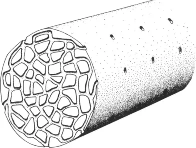
Fig. 1 Drawing of a muscle fiber cut across through one T system network. The openings of other T system networks are seen along the length of the muscle fiber. Not all tubules near the sur...
Table of contents
- Cover image
- Title page
- Table of Contents
- CONTRIBUTORS
- Copyright
- LIST OF CONTRIBUTORS
- PREFACE
- PREFACE TO THE FIRST EDITION
- CONTENTS OF OTHER VOLUMES
- Chapter 1: ELECTRICAL PROPERTIES OF THE TRANSVERSE TUBULAR SYSTEM
- Chapter 2: THE NEUROMUSCULAR JUNCTION—THE ROLE OF ACETYLCHOLINE IN EXCITABLE MEMBRANES
- Chapter 3: SOME ASPECTS OF THE BIOPHYSICS OF MUSCLE
- Chapter 4: ENERGY NEED, DELIVERY, AND UTILIZATION IN MUSCULAR EXERCISE
- Chapter 5: THE CONTROL OF MUSCULAR ACTIVITY BY THE CENTRAL NERVOUS SYSTEM
- Chapter 6: ELECTROMYOGRAPHY
- Chapter 7: PROTEINS OF THE MYOFIBRIL
- Chapter 8: BIOCHEMISTRY OF MUSCLE
- Chapter 9: BIOCHEMISTRY OF MUSCLE MITOCHONDRIA
- Chapter 10: ATP BREAKDOWN FOLLOWING ACTIVATION OF MUSCLE
- AUTHOR INDEX
- SUBJECT INDEX
Frequently asked questions
Yes, you can cancel anytime from the Subscription tab in your account settings on the Perlego website. Your subscription will stay active until the end of your current billing period. Learn how to cancel your subscription
No, books cannot be downloaded as external files, such as PDFs, for use outside of Perlego. However, you can download books within the Perlego app for offline reading on mobile or tablet. Learn how to download books offline
Perlego offers two plans: Essential and Complete
- Essential is ideal for learners and professionals who enjoy exploring a wide range of subjects. Access the Essential Library with 800,000+ trusted titles and best-sellers across business, personal growth, and the humanities. Includes unlimited reading time and Standard Read Aloud voice.
- Complete: Perfect for advanced learners and researchers needing full, unrestricted access. Unlock 1.4M+ books across hundreds of subjects, including academic and specialized titles. The Complete Plan also includes advanced features like Premium Read Aloud and Research Assistant.
We are an online textbook subscription service, where you can get access to an entire online library for less than the price of a single book per month. With over 1 million books across 990+ topics, we’ve got you covered! Learn about our mission
Look out for the read-aloud symbol on your next book to see if you can listen to it. The read-aloud tool reads text aloud for you, highlighting the text as it is being read. You can pause it, speed it up and slow it down. Learn more about Read Aloud
Yes! You can use the Perlego app on both iOS and Android devices to read anytime, anywhere — even offline. Perfect for commutes or when you’re on the go.
Please note we cannot support devices running on iOS 13 and Android 7 or earlier. Learn more about using the app
Please note we cannot support devices running on iOS 13 and Android 7 or earlier. Learn more about using the app
Yes, you can access Physiology and Biochemistry by Geoffrey Bourne in PDF and/or ePUB format, as well as other popular books in Social Sciences & Physical Anthropology. We have over one million books available in our catalogue for you to explore.