eBook - ePub
Magnetic Resonance in Epilepsy
Neuroimaging Techniques, Second Edition
- 464 pages
- English
- ePUB (mobile friendly)
- Available on iOS & Android
eBook - ePub
Magnetic Resonance in Epilepsy
Neuroimaging Techniques, Second Edition
About this book
Remarkable advances in imaging have increased the importance of MRI for diagnostic, treatment and management of epilepsy. Neuroimaging of patients with epilepsy no longer simply deals with the technology and interpretation of images but also with issues of brain metabolism, energetics, cognition and brain dysfunction. The first edition of Magnetic Resonance in Epilepsy came into clinical practice in 1995 with a revolutionary idea; that is, MR is as important as EEG in the clinical management of patients with epilepsy. The second edition of Magnetic Resonance in Epilepsy, the only comprehensive text in the field of epilepsy neuroimaging, reviews fundamental concepts and new advances in MR technology, computerized analysis, MR spectroscopy, DWI and other neuroimaging techniques such as PET, SPECT and MEG application to the study of patients with epileptic disorders.*Provides a crucial update of recent advances in imaging techniques*Timely publication as subject of neuroimaging is a very "hot" area in both clinical epilepsy and basic neuroscience research*Editors are well-respected in this field
Trusted by 375,005 students
Access to over 1.5 million titles for a fair monthly price.
Study more efficiently using our study tools.
Information
CHAPTER 1
Introduction to Epilepsy
Graeme D. Jackson, Ruben I. Kuzniecky and Samuel F. Berkovic
“Today, epilepsy has more secrets to confide to the neurologist or neurosurgeon who can understand the ‘tongue’ she speaks.”
Wilder Penfield, 1974 (age 83).
The origin of the modern view of epilepsy is generally considered to have been the studies and work of John Hughlings Jackson (Fig. 1.1). He was appointed assistant physician to the National Hospital for the Relief and Cure of the Paralysed and Epileptic (now the National Hospital for Neurology and Neurosurgery), Queen Square, London in 1862 at the age of 27. Jackson’s wife developed focal motor seizures following a cerebral thrombosis, and it is this form of epilepsy, with its typical march of symptoms, that became known as jacksonian epilepsy. This work was in the context of a developing view of neurology that was beginning to relate cortical functions to specific locations within the brain (1). In 1873 Jackson presented his classic definition of epileptic seizures, which was ‘occasional sudden excessive, rapid and local discharges of gray matter (2, 3).’ This view established partial seizures as being truly epileptic in origin. At that time William Gowers, J. Hughlings Jackson, Victor Horsley, and David Ferrier were developing concepts of cortical localization and of epilepsy, which were to have far reaching consequences. The combination of basic scientific research and clinical findings enabled Jackson’s ideas to be confirmed by Ferrier in experiments in primates using techniques of cortical stimulation (4). As a consequence, brain functions were able to be localized and the view developed that localization of the site of origin of seizures was possible based on their clinical symptoms–in particular the symptoms at the start of the clinical seizure.
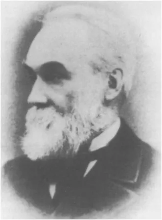
FIG. 1.1 John Hughlings Jackson, London (1835–1911).
ORIGINS OF SEIZURE SURGERY
Using this concept of brain localization and using clinical symptoms as a guide to the site of the seizure focus (jacksonian seizures), a neurosurgeon in Glasgow, William Macewen, correctly localized a frontal meningioma and operated in 1879 (5). Following tumor removal the patient both survived and became seizure-free. At the National Hospital in London, Victor Horsley operated on three patients with epileptogenic lesions in 1886 (6). The first of these was a patient of Jackson’s, and present in the operating theater were Horsley, Jackson, and Ferrier. This represents the origin of epilepsy surgery for the treatment of epilepsy. Although these early cases were successful, establishing the principle of seizure control by surgical removal of the ‘seizure focus,’ the overall success of surgery for epilepsy was poor and eventually operative treatment fell into disuse until the ‘modern era’ represented by Penfield and colleagues at the Montreal Neurologic Institute. What now seem like simple measures, such as anesthesia, antisepsis, and antibiotics, made a large impact on the feasibility and safety of neurosurgery for epilepsy, as they did in many other areas of medicine.
Despite major developments in the understanding of the process of epilepsy and the propagation and spread of seizures, the central issues and principles on which this surgery was based have, at their core, remained largely unchanged to the present day. That is, if the seizure focus can be accurately localized, then surgical removal of that focus will alleviate the occurrence of seizures. What has changed to a remarkable extent is the technological environment in which this work can be carried out. Major milestones have been the advent of the electroencephalogram (EEG) and more recently neuroimaging techniques, represented in their most dramatic form by magnetic resonance imaging (MRI). The data and criteria by which the diagnosis of seizure localization is made continues to develop and this book examines the role of MR in this evaluation process: this development has changed the face of epilepsy diagnosis, management and treatment. In addition to its initial revolutionary role in surgical treatment of epilepsy, MRI has altered the understanding of, and clinical approach to, many forms of focal and generalized epi1epsy.
THE MODERN ERA
During the 20th century, Wilder Penfield (Fig. 1.2), who founded the Montreal Neurologic Institute in 1934, became the leading figure in the further development of both the surgical treatment of epilepsy and the structural localization of brain functions. In common with Jackson, his extensive writings, coupled with a clear, detailed, and objective skill in observation, provided a wealth of information, which is of relevance to this day. From his observations, Penfield recognized the dramatic vascular changes that occur during seizures (7). Despite the subsequent emphasis, as a consequence of the development of the powerful EEG technology, on the electrical events that occur during seizures, Penfield retained his interest in these vascular changes throughout his life (8). With the advent of ictal imaging technologies, such as SPECT (9) and functional MRI, the legacy of his observations remain relevant to the interpretation of blood flow changes during ictal events. Although he died in 1976 at the age of 85, before the development of medical nuclear magnetic resonance technology, he clearly foresaw that new technology would reveal many new aspects of epilepsy and brain functions.

FIG. 1.2 Wilder Penfield, Montreal (1891–1976).
ELECTROENCEPHALOGRAM DEVELOPMENT
In 1929, Berger published the technique of EEG (10), which, in his study, was based on a single-electrode contact. This important technology was subsequently developed by many researchers to provide a means whereby seizures could be localized without characteristic and localizing ictal clinical symptoms. This was a very exciting development in neurology as it enabled the totally noninvasive investigation of brain function with an objective technique that provided information independent of the clinical examination. This eventually shifted the emphasis from epilepsy originating in eloquent areas of cortex such as the motor strip (because of the ability to localize these events) and enabled temporal lobe seizures to be localized on the basis of the electroencephalographic localization of the electrical activity associated with the seizure.
The EEG technology developed to the point that in 1951 Bailey (neurosurgeon) and Gibbs (electroencephalographer) published a series of 25 patients who underwent temporal lobe resection based only on EEG criteria (11) without supporting clinical localization. The results were encouraging and helped establish the EEG as the prime method of seizure localization, to the extent that epileptic syndromes often became referred to as electro-clinical diagnoses (showing how important this investigation modality had become in the practice of epilepsy). Over the next 30 years the study of epilepsy was dominated by the technical development of, and understanding of the information obtained from, the EEG (12, 13). During this period, surgery, as a treatment of epilepsy, also gained considerable ground.
MAGNETIC RESONANCE IMAGING
Magnetic resonance techniques were first applied to human studies in the late 1970s (12) and the routine clinical availability of MRI began in the mid 1980s. MRI enables a detailed view of the fine structure of the living brain, which had previously only been possible after death (by postmortem examination). It soon became apparent that MRI could add another powerful dimension to the problem of localizing where seizures originate by demonstrating the location and nature of structural brain abnormalities. Small tumors, many which had been undetected by CT scanning, were commonly found in these patients (13–17).
Less apparent, but especially important, lesions such as hippocampal sclerosis were initially thought to be largely undetectable using MR (14, 16, 18). The development of MR techniques for the detection of hippocampal sclerosis figured prominently in the first edition of this book and remains central t...
Table of contents
- Cover image
- Title page
- Table of Contents
- Inside Front Cover
- Fornt Matter
- Copyright
- Dedication
- List of Contributors
- Editors
- Foreword
- Preface to the Second Edition
- Acknowledgments
- Chapter 1: Introduction to Epilepsy
- Chapter 2: Principles of Magnetic Resonance Imaging
- Chapter 3: Brain Anatomy
- Chapter 4: Temporal Lobe Epilepsy
- Chapter 5: Extra-Temporal Lobe Epilepsy
- Chapter 6: MRI in Special Conditions Associated with Epilepsy
- Chapter 7: Malformations of Cortical Development
- Chapter 8: Structural Analysis Applied to Epilepsy
- Chapter 9: Imaging and Neuropsychology
- Chapter 10: Functional MRI in Epilepsy
- Chapter 11: Magnetic Resonance Neurophysiology: Simultaneous EEG and fMRI
- Chapter 12: MR Diffusion and Perfusion Imaging in Epilepsy
- Chapter 13: Magnetic Resonance Spectroscopy
- Chapter 14: Single-photon-emission Computed Tomography in Epilepsy
- Chapter 15: Positron-emission Tomography in Epilepsy
- Chapter 16: Magnetoencephalography in Epilepsy
- Index
Frequently asked questions
Yes, you can cancel anytime from the Subscription tab in your account settings on the Perlego website. Your subscription will stay active until the end of your current billing period. Learn how to cancel your subscription
No, books cannot be downloaded as external files, such as PDFs, for use outside of Perlego. However, you can download books within the Perlego app for offline reading on mobile or tablet. Learn how to download books offline
Perlego offers two plans: Essential and Complete
- Essential is ideal for learners and professionals who enjoy exploring a wide range of subjects. Access the Essential Library with 800,000+ trusted titles and best-sellers across business, personal growth, and the humanities. Includes unlimited reading time and Standard Read Aloud voice.
- Complete: Perfect for advanced learners and researchers needing full, unrestricted access. Unlock 1.5M+ books across hundreds of subjects, including academic and specialized titles. The Complete Plan also includes advanced features like Premium Read Aloud and Research Assistant.
We are an online textbook subscription service, where you can get access to an entire online library for less than the price of a single book per month. With over 1.5 million books across 990+ topics, we’ve got you covered! Learn about our mission
Look out for the read-aloud symbol on your next book to see if you can listen to it. The read-aloud tool reads text aloud for you, highlighting the text as it is being read. You can pause it, speed it up and slow it down. Learn more about Read Aloud
Yes! You can use the Perlego app on both iOS and Android devices to read anytime, anywhere — even offline. Perfect for commutes or when you’re on the go.
Please note we cannot support devices running on iOS 13 and Android 7 or earlier. Learn more about using the app
Please note we cannot support devices running on iOS 13 and Android 7 or earlier. Learn more about using the app
Yes, you can access Magnetic Resonance in Epilepsy by Ruben Kuzniecky,Graeme D. Jackson in PDF and/or ePUB format, as well as other popular books in Medicine & Neurology. We have over 1.5 million books available in our catalogue for you to explore.