Translational Neuroimaging
eBook - ePub

Translational Neuroimaging

Tools for CNS Drug Discovery, Development and Treatment

  1. 464 pages
  2. English
  3. ePUB (mobile friendly)
  4. Available on iOS & Android
eBook - ePub

Translational Neuroimaging

Tools for CNS Drug Discovery, Development and Treatment

Book details
Book preview
Table of contents
Citations

About This Book

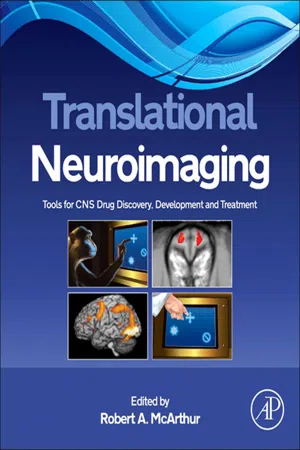
Translational Neuroimaging: Tools for CNS Drug Discovery, Development and Treatment combines the experience of academic, clinical and industrial neuroimagers in a unique collaborative approach to provide an integrated perspective of the use of small animal and human brain imaging in developing and validating translational models and biomarkers for the study and treatment of neuropsychiatric disorders. Translational Neuroimaging: Tools for CNS Drug Discovery, Development and Treatment examines the translational role of neuroimaging in model development from preclinical animal models, to human experimental medicine, and finally to clinical studies. The focus of this book is to identify and provide common endpoints between species that can serve to inform both the clinic and the bench with the information needed to accelerate clinically-effective CNS drug discovery. This book covers methodical issues in human and animal neuroimaging translational research as well as detailed applied examples of the use of neuroimaging in neuropsychiatric disorders and the development of drugs for their treatment. Translational Neuroimaging: Tools for CNS Drug Discovery, Development and Treatment appeals to non-clinical and clinical neuroscientists working in and studying neuropsychiatric disorders and their treatment as well as providing the novice researcher or researcher outside of his/her expertise the opportunity to understand the background of translational research and the use of imaging in this field.

  • Provides a background to translational research and the use of brain imaging in neuropsychiatric disorders
  • Critical discussion of the potential and limitations of neuroimaging as a translational tool for identifying and validating biomarkers
  • Identifies cross species neurosystems and common endpoints necessary to help accelerate CNS drug discovery and development for the treatment of neuropsychiatric disorders

Frequently asked questions

Simply head over to the account section in settings and click on “Cancel Subscription” - it’s as simple as that. After you cancel, your membership will stay active for the remainder of the time you’ve paid for. Learn more here.
At the moment all of our mobile-responsive ePub books are available to download via the app. Most of our PDFs are also available to download and we're working on making the final remaining ones downloadable now. Learn more here.
Both plans give you full access to the library and all of Perlego’s features. The only differences are the price and subscription period: With the annual plan you’ll save around 30% compared to 12 months on the monthly plan.
We are an online textbook subscription service, where you can get access to an entire online library for less than the price of a single book per month. With over 1 million books across 1000+ topics, we’ve got you covered! Learn more here.
Look out for the read-aloud symbol on your next book to see if you can listen to it. The read-aloud tool reads text aloud for you, highlighting the text as it is being read. You can pause it, speed it up and slow it down. Learn more here.
Yes, you can access Translational Neuroimaging by Robert A. McArthur in PDF and/or ePUB format, as well as other popular books in Médecine & Neurologie. We have over one million books available in our catalogue for you to explore.

Information

Year
2012
ISBN
9780123869975
Subtopic
Neurologie

Chapter 1

Neuroimaging Modalities
Description, Comparisons, Strengths, and Weaknesses

Richard G. Wise
Cardiff University Brain Research Imaging Centre (CUBRIC), School of Psychology, Cardiff University, Park Place, Cardiff CF10 3AT, United Kingdom
1.0. Introduction
1.1. Windows to the Brain
1.1.1. Brain Structure
1.1.2. Brain Function
2.0. Radiotracer Techniques
2.1. Single-Photon Emission Computed Tomography
2.2. Positron Emission Tomography
2.2.1. Cerebral Metabolism and Blood Flow
2.2.2. Receptor Studies
3.0. Electrophysiological Techniques
3.1. Electroencephalography
3.2. Magnetoencephalography
4.0. Magnetic Resonance Techniques
4.1. MRS
4.2. Structural MRI
4.3. fMRI
4.3.1. BOLD fMRI
4.3.2. Pharmacological fMRI
4.3.3. Arterial Spin Labeling fMRI
5.0. Advantages, Disadvantages, and Practical Considerations
Summary
In this chapter, I review the most important neuroimaging methods that are gaining ground as translational tools in central nervous system (CNS) drug discovery, drug development and treatment. I consider the information that may be gained about brain structure and brain function from each of the techniques, focusing in particular on positron emission tomography (PET), magnetic resonance imaging (MRI), magnetic resonance spectroscopy (MRS), electroencephalography (EEG) and magnetoencephalography (MEG). I explore the signals that they provide and how they may be used, along with their relative advantages, disadvantages, and the practicalities of using the different techniques. This roadmap through the range of the most widely applied neuroimaging techniques relevant to translational research provides an introduction for the later chapters that describe applications in specific disease areas.

1.0 Introduction

This chapter aims to provide an introduction to the neuroimaging modalities that are important to drug discovery, development and treatment. I describe the basis of each technique and compare each one with the others, offering a roadmap through the jungle of different types of neuroimaging, including in particular magnetic resonance (MR) techniques, positron emission tomography (PET), and electrophysiological measurements. I will describe the nature of each measurement or imaging signal and the functional readout from the biology, as well as what they are used for and how they are applied. I discuss the strengths and weaknesses of the different techniques, as one modality rarely has the capability of answering all of the relevant questions. I discuss principally the application of the neuroimaging modalities common to animals and humans, focusing in particular on noninvasive techniques.
I begin the chapter with an overview of the biological domains overseen by the different imaging techniques before describing each modality in turn. I then discuss the advantages and disadvantages of the modalities.

1.1 Windows to the Brain

There is no single neuroimaging modality that can offer the complete picture of brain structure or function that is needed for drug development and assessing disease or treatment effects. Each modality has its own sensitivity profile that may offer a piece of information directly linked to the action of a pharmacological agent, e.g. receptor binding, or more likely an indirect marker of pharmacological or disease action, e.g. local hemodynamic activity. It is necessary to appreciate the basis of the neuroimaging signals and their limitations in order to interpret them. This is particularly important in the process of decision making in drug discovery and development. Over- or misinterpretation of neuroimaging data could lead to the expensive failure of new compounds late in the drug development process, which could have been anticipated earlier. Underuse of valuable information contained in neuroimaging data might lead to promising compounds being abandoned unnecessarily.
The neuroimaging modalities applied in humans and animals may be categorized as offering either a structural or a functional readout. While often a useful distinction, the boundary between structure and function may be blurred, in particular at the micro scale and where function and structure are intimately linked. An elegant example lies in the characterization of the diffusion and distribution of water in the human brain. Diffusion-based MR imaging is regarded as offering structural information. However, in the context of stroke, where there is a redistribution of intra- and extracellular water, structural information and functional viability of tissue become closely linked. Furthermore, the diffusion of water in the brain has been demonstrated to change over the comparatively short timescales associated with structural plasticity during learning.1 In more general terms, the function of a neuron might be said to be defined by its connections, local and remote, to other neurons, such connections being identified noninvasively by diffusion tractography techniques in white matter.

1.1.1 Brain Structure

Alterations in brain structure with disease or treatment range from the scale of gross structure, including regional tissue volumes, through to micro and molecular structure, such as receptor distributions. Techniques sensitive to alterations in brain structure most commonly offer a long-term marker of disease progression or modification with treatment. This is because macro-scale alterations in structure, e.g. brain atrophy, normally develop over long periods, typically years in humans.
Structural magnetic resonance imaging (MRI) techniques are particularly suited to examining long-term structural alterations because of their noninvasiveness and therefore their ability to follow longitudinal changes in a research cohort. Traditional MRI structural markers, such as T1 and T2 relaxation time constants,i can reveal fat and water distribution (as well as other physicochemical differences), thus distinguishing gray matter, white matter, and cerebrospinal fluid, while developments in MR image contrast, such as magnetization transfer, can offer information on pools of liquid and macromolecular water with potential uses in examining demyelinating conditions.2 Microscopy is invasive and so only available post mortem, but is useful for characterizing altered brain microstructure. Microstructural alterations at the scale of molecular receptors can be probed with radiotracers using PET, looking for example, at alterations in spatial distributions of specific receptors. Such molecular-level alterations are often interpreted as having direct functional consequences.

1.1.2 Brain Function

Brain function is characterized by many different activities of the brain accessible to neuroimaging techniques. Functional processes are normally those occurring over short timescales from small fractions of a second to minutes. They support or are associated with information processing or the transmission and reception of signals in the brain. In animals, it is possible to place electrodes within brain tissue and to record individual cellular potentials or the electrical activity of groups of cells. In humans, this is only possible in rare circumstances when electrodes are implanted for reasons of surgical diagnosis, for example to identify seizure foci or brain stimulation. More commonly, we rely on indirect or ensemble measures of brain function, which may either be focused on specific portions of brain tissue or distributed across the brain to study the brain at the systems level.
The brain’s neuronal activity is linked or coupled to its blood supply,3,4 allowing altered hemodynamics and therefore regional blood oxygenation to be used as a marker of altered function. The growth of functional neuroimaging studies in humans in the last 20 years has exploited these phenomena, first with PET and more recently and on a larger scale with functional magnetic resonance imaging (fMRI). Optical imaging techniques, including invasive cortical infrared imaging and noninvasive near-infrared spectroscopy (NIRS) rely on changes in cerebral blood oxygenation.
A continuous energy supply to brain tissue is essential for maintaining ion concentration gradients and therefore electrical potentials. Some neuroimaging techniques have been developed that are sensitive to alterations in cerebral metabolism and, in particular, the biochemical species involved in energy supply. PET can be made sensitive to oxygen or glucose metabolism. fMRI techniques are also emerging that are able to quantify cerebral oxygen consumption. Magnetic resonance spectroscopy (MRS) can measure chemical concentrations within brain tissue and therefore monitor the energy status of tissue through species such as high-energy phosphates. With the appropriate use of tracers detectable with MRS, rate constants and chemical fluxes can be estimated to quantify cerebral metabolism.5
Specific molecules engaged in signaling or processes associated with synaptic transmission can be studied using PET and MRS. With the development of appropriate PET ligands, specific receptor activity and the distribution of receptors can now be assessed. Receptor activity has the advantage of being directly associable with the action of pharmacological agents in the brain, whereas MRS is able to measure the bulk concentration of the more common neurotransmitters and their modulation with disease and pharmacological intervention. It must be borne in mind, however, that the relationship between neurotransmitter concentration and brain function may be a complex one depending on the availability or otherwise of the neurotransmitter.
Noninvasive measures of electrophysiological activity can be made from the scalp by recording electrical potentials (i.e. electroencephalography; EEG) or the tiny magnetic fields associated with neuronal activity (i.e. magnetoencephalography; MEG). In order to be detectable at a distance of centimeters from their source, these signals necessarily arise from the coordinated activity of populations of neurons.
2.0 Radiotracer Techniques

Radiotracer techniques use radionuclides as probes to quantify physiological processes, e.g. cerebral blood flow, or to label biochemical pathways or specific molecules. The probe is spatially localized by detecting the emitted radioactivity, while variations in local radioactivity over time can be used to identify rate constants and physiological fluxes. Radiotracers are sensitive and can be adapted to different uses, including marking substrates to investigate biochemical processes, labeling a drug target, or labeling the drug itself. Molecular tracers are the only way to measure receptors and their function because of their low concentrations (nano- and picomolar range).

2.1 Single-Photon Emission Computed Tomography

Single-photon emission computed tomography (SPECT) detects tracer molecules labeled with gamma-emitting radioisotopes. Typically it uses an array of two or three gamma cameras that rotate around the subject and is increasingly combined with a computed tomography (CT) system that offers improved spatial resolution and registration with structural images. SPECT is more widely available than PET, although the range of tracers for SPECT is more limited.
SPECT is commonly used for measuring cerebral perfusion (regional cerebral...

Table of contents

  1. Cover image
  2. Title page
  3. Table of Contents
  4. Copyright
  5. Dedication
  6. Preface
  7. Contributors
  8. Acknowledgments
  9. Abbreviation List
  10. Chapter 1. Neuroimaging Modalities: Description, Comparisons, Strengths, and Weaknesses
  11. Chapter 2. Magnetic Resonance Imaging as a Tool for Modeling Drug Treatment of CNS Disorders: Strengths and Weaknesses
  12. Chapter 3. Small Animal Imaging as a Tool for Modeling CNS Disorders: Strengths and Weaknesses
  13. Chapter 4. Structural Magnetic Resonance Imaging as a Biomarker for the Diagnosis, Progression, and Treatment of Alzheimer Disease
  14. Chapter 5. Positron Emission Tomography in Alzheimer Disease: Diagnosis and Use as Biomarker Endpoints
  15. Chapter 6. Rethinking the Contribution of Neuroimaging to Translation in Schizophrenia
  16. Chapter 7. Neuroimaging as a Translational Tool in Animal and Human Models of Schizophrenia
  17. Chapter 8. Functional Magnetic Resonance Imaging as a Biomarker for the Diagnosis, Progression, and Treatment of Autistic Spectrum Disorders
  18. Chapter 9. Translational Neuroimaging for Drug Discovery and Development in Autism Spectrum Disorders: Guidance from Clinical Imaging and Preclinical Research
  19. Chapter 10. Neuroimaging as a Biomarker for the Diagnosis, Progression, and Treatment of Substance Abuse Disorders
  20. Chapter 11. Translational Neuroimaging: Substance Abuse Disorders
  21. Chapter 12. Neuroimaging Approaches to the Understanding of Depression and the Identification of Novel Antidepressants
  22. Index