eBook - ePub
Essential Clinically Applied Anatomy of the Peripheral Nervous System in the Head and Neck
- 202 pages
- English
- ePUB (mobile friendly)
- Available on iOS & Android
eBook - ePub
Essential Clinically Applied Anatomy of the Peripheral Nervous System in the Head and Neck
About this book
Essential Clinically Applied Anatomy of the Nerves in the Head and Neck presents the reader with an easy access format to clinically-applied peripheral nervous system (PNS) anatomy. Perfect for a quick reference to essential details. The chapters review nerves of the head and neck, the origin(s), course, distribution and relevant pathologies affecting each are given, where relevant. The pathologies present typical injuries to the nerves of the PNS, as well as clinical findings on examination and treatments. It details modern clinical approaches to the surgery and other treatments of these nerve pathologies, as applicable to the clinical scenario.
- Surveys the anatomy of the PNS nerves in the head and neck
- Includes key facts and summary tables essential to clinical practice
- Offers a succinct yet comprehensive format with quick and easy access to facts and essential details
- Includes comprehensive chapters on nerves of the head and neck, discussing origin, course, distribution, and relevant pathologies
Trusted by 375,005 students
Access to over 1.5 million titles for a fair monthly price.
Study more efficiently using our study tools.
Information
Topic
Ciencias biológicasSubtopic
NeurologíaChapter 1
Overview of the Nervous System
Abstract
This chapter will examine the nervous system providing an overview of the key components from its divisions. There are two main ways to examine the nervous system—from a structural viewpoint and also from a functional perspective. It will highlight the main areas of the brain and spinal cord, as well as the basic constituents of the cells of the nervous system. It introduces the concept of the sympathetic and parasympathetic nervous system and the major roles arts of the nervous system have.
Keywords
Central nervous system; peripheral nervous system; spinal cord; somatic nervous system; autonomic nervous system; functional division
1.1 Overview of the Nervous System
Broadly speaking, the nervous system is divided into two components—central and peripheral. The central nervous system (CNS) comprises the brain as well as the spinal cord. The peripheral nervous system (PNS) comprises all of the nerves—cranial, spinal, and peripheral nerves, including the sensory and motor nerve endings of these nerves.
1.2 Divisions of the Nervous System
1.2.1 Central Nervous System
The CNS is comprised of the brain as well as the spinal cord. The purpose of the CNS is to integrate all the body functions from the information it receives. Within the PNS, there are many, many nerves (group of nerve fibers together), however, the CNS does not contain nerves. Within the CNS, a group of nerve fibers traveling together is called a pathway, or tract. If it links the left and right hand sides it is referred to as a commissure.
1.2.1.1 Neurons
Within the CNS, there are many, many millions of nerve cells called neurons. Neurons are cells which are electrically excitable and transmit information from one neuron to another by chemical and electrical signals. There are three very broad classifications of neurons—sensory (which process information on light, touch, and sound to name some of the modalities), motor (supplying muscles), and interneurons (which interconnect neurons via a network).
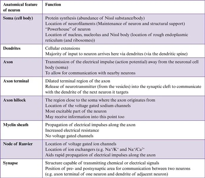
Typically a neuron comprises some basic features, however there are a variety of specializations that some have dependent on the location within the nervous system. In general, a neuron has a cell body. Here, the nucleus—the powerhouse—of the neuron lies with its cytoplasm. At this point, numerous fine fibers enter called dendrites. These processes receive information from adjacent neurons keeping it up-to-date with the surrounding environment. This way the amount of information that a single neuron receives is significantly increased. From a neuron, there is a long single process of variable length called an axon. This conducts information away from the neuron. Some neurons however have no axons and the dendrites will conduct information to and from the neuron. In addition to this, a lipoprotein layer called the myelin sheath can surround the axon of a principal cell. This is not a continuous layer along the full length of the axon. Rather, there are interruptions called nodes of Ranvier. It is at this point where the voltage gated channels occur, and it is at that point where conduction occurs. Therefore, the purpose of the myelin sheath is to enable almost immediate conduction between one node of Ranvier and the next ensuring quick communication between neurons.
In relation to the size of neurons, this varies considerably. The smallest of neurons can be as small as 5 μm, with the largest for example motor neurons, can be as big as 135 μm. In addition, axonal length can vary considerably too. The shortest of these can be 100 μm, whereas a motor axon supplying the lower limb for example the toes, can be as long as 1 m.
In the PNS, neurons are found in ganglia, or in laminae (layers) or nuclei in the CNS.
Neurons communicate with each other at a point called a synapse. Most of these junctional points are chemical synapses where there is the release of a neurotransmitter which diffuses across the space between the two neurons. The other type of synapse is called an electrical synapse. This form is generally more common in the invertebrates, where there is close apposition of one cell membrane and the next that is at the pre- and postsynaptic membranes. Linking these two cells is a collection of tubules called connexons. The transmission of impulses occurs in both directions and very quickly. This is because there is no delay in the neurotransmitter having to be activated and released across the synapse. Instead, the flow of communication depends on the membrane potentials of the adjacent cells (Table 1.1).
Table 1.1
This Summarizes the Main Cellular Components of Nervous Tissue, Including the Role(s) for These
1.2.1.2 Neuroglia
Neuroglia, or glia, are the supportive cells for neurons. Their main purpose is not in relation to the transmission of nerve impulses. Rather, they are involved in providing nutrient support, maintenance of homeostasis, and the production of the myelin sheath. There are two broad classifications—microglia and macroglia.
The microglia have a defence role as a phagocytic cell. They are found throughout the brain and spinal cord, and can alter their shape, especially when they engulf particulate material. They are thus serving a protective role for the nervous system.
Macroglia are subdivided into seven different types, again with each having a special role.
1. Astrocytes
These cells fill in the spaces between neurons and provide for structural integrity. They also have processes which join to the capillary blood vessels. These are known as perivascular end feet. Therefore, with their close apposition to blood vessels, they are also thought to be responsible for metabolite exchange between the neurons and the vasculature. They are found in the CNS.
These cells fill in the spaces between neurons and provide for structural integrity. They also have processes which join to the capillary blood vessels. These are known as perivascular end feet. Therefore, with their close apposition to blood vessels, they are also thought to be responsible for metabolite exchange between the neurons and the vasculature. They are found in the CNS.
2. Ependymal cells
There are three types of ependymal cells—ependymocytes, tanycytes, and choroidal epithelial cells. The ependymocytes allow for the free movement of molecules between the cerebrospinal fluid (CSF) and the neurons. Tanycytes are generally found in the third ventricle and can be involved in responding to changing hormonal levels of the blood derived hormones in the CSF. Choroidal epithelial cells are the cells which control the chemical composition of the CSF. They are found in the CNS.
There are three types of ependymal cells—ependymocytes, tanycytes, and choroidal epithelial cells. The ependymocytes allow for the free movement of molecules between the cerebrospinal fluid (CSF) and the neurons. Tanycytes are generally found in the third ventricle and can be involved in responding to changing hormonal levels of the blood derived hormones in the CSF. Choroidal epithelial cells are the cells which control the chemical composition of the CSF. They are found in the CNS.
3. Oligodendrocytes
These cells are responsible for the production of myelin sheaths. They are found in the CNS.
These cells are responsible for the production of myelin sheaths. They are found in the CNS.
4. Schwann cells
Like oligodendrocytes, Schwann cells are responsible for the production of the myelin sheath, but in the PNS. They also have an additional role in phagocytosis of any debris; therefore help to clean the surrounding environment.
Like oligodendrocytes, Schwann cells are responsible for the production of the myelin sheath, but in the PNS. They also have an additional role in phagocytosis of any debris; therefore help to clean the surrounding environment.
5. Satellite cells
These cells surround those neurons of the autonomic system and also the sensory system. They maintain a stable chemical balance of the surrounding environment to the neurons. They are therefore found in the PNS.
These cells surround those neurons of the autonomic system and also the sensory system. They maintain a stable chemical balance of the surrounding environment to the neurons. They are therefore found in the PNS.
6. Radial glia
Radial glial cells act as scaffolding onto which new neurons migrate to. They are found in the CNS.
Radial glial cells act as scaffolding onto which new neurons migrate to. They are found in the CNS.
7. Enteric glia
These cells are found within the gastrointestinal tract and aid digestion and maintenance of homeostasis. They are by their very nature found in the PNS.
These cells are found within the gastrointestinal tract and aid digestion and maintenance of homeostasis. They are by their very nature found in the PNS.
1.2.1.3 Gray and White Matter
In the CNS, there are two clear differences between the structural components. It is divided by its appearance of either gray or white matter. Within the gray matter there are cell bodies and dendrites of efferent neurons, glial cells (supportive), fibers of afferent neurons and interneurons. The white matter on the other hand primarily consists of myelinated axons and the supportive glial cells. The purpose of the white matter is to allow for communication from one part of the cerebrum to the other, and also to communicate to other brain areas and carry impulses through the spinal cord.
1.2.1.4 Brain
The brain is comprised of three swellings which form during development—the forebrain (prosencephalon), midbrain (mesencephalon), and hindbrain (rhombencephalon). During development in mammals, the forebrain continues to grow, whereas in other vertebrates for example amphibians and fish, the three divisions remain in proportion to each other during growth.
The brain can also be subdivided into the following:
a. Telencephalon (cerebral hemispheres)+Diencephalon (thalamus and hypothalamus)=FOREBRAIN
b. Mesencephalon=MIDBRAIN
c. Metencephalon (pons, cerebellum, and the trigeminal, abducent, facial, and vestibulocochlear nerves)+Myelencephalon (medulla oblongata)=HINDBRAIN
Surrounding the core of the forebrain that is the diencephalon are the two large cerebral hemispheres (left and right), which constitute the cerebrum. The cerebrum is composed of three regions:
1. Cerebral cortex
The cerebral cortex is the gray matter of the cerebrum. It is comprised of three parts based on its functions—motor, sensory, and association areas. The motor area is present in both cerebral cortices. Each one controls the opposite side of the body that is the left motor area controls the right side of the body, and vice versa. There are two broad regions—a primary motor area responsible for execution of voluntary movements, and supplementary area involved in selection of voluntary movements.
The sensory area receives information from the opposite side of the body ie, the right cerebral cortex receives sensory information from the left side of the body. In essence it deals with auditory information (via the primary auditory cortex), visual information (via the primary visual cortex), and sensory information (via the primary somatosensory cortex).
The association areas allow us to understand the external environment. All of the cerebral cortex is subdivided into lobes of the brain. These are:
a. Frontal lobes
Broadly speaking the frontal lobe deals with “executive” functions and our long-term memory. It also is the site of our primary motor cortex, toward its posterior part.
Broadly speaking the frontal lobe deals with “executive” functions and our long-term memory. It also is the site of our primary motor cortex, toward its posterior part.
b. Parietal lobes
The parietal lobes are responsible for integration of sensory functions. It is the site of our primary somatosensory cortex.
The parietal lobes are responsible for integration of sensory functions. It is the site of our primary somatosensory cortex.
c. Temporal lobes
The temporal lobes integrate information related to hearing, and therefore, is the site of our primary auditory cortex.
The temporal lobes integrate information related to hearing, and therefore, is the site of our primary auditory cortex.
d. Occipital lobes
The occipital lobes integrate our visual information and function as the primary visual cortex.
The occipital lobes integrate our visual information and function as the primary visual cortex.
2. Basal ganglia
The basal ganglia are three sets of nuclei—the globus pallidus, str...
The basal ganglia are three sets of nuclei—the globus pallidus, str...
Table of contents
- Cover image
- Title page
- Table of Contents
- Copyright
- List of Figures
- List of Tables
- Preface
- Acknowledgments
- Chapter 1. Overview of the Nervous System
- Chapter 2. Head
- Chapter 3. Neck
- Index
Frequently asked questions
Yes, you can cancel anytime from the Subscription tab in your account settings on the Perlego website. Your subscription will stay active until the end of your current billing period. Learn how to cancel your subscription
No, books cannot be downloaded as external files, such as PDFs, for use outside of Perlego. However, you can download books within the Perlego app for offline reading on mobile or tablet. Learn how to download books offline
Perlego offers two plans: Essential and Complete
- Essential is ideal for learners and professionals who enjoy exploring a wide range of subjects. Access the Essential Library with 800,000+ trusted titles and best-sellers across business, personal growth, and the humanities. Includes unlimited reading time and Standard Read Aloud voice.
- Complete: Perfect for advanced learners and researchers needing full, unrestricted access. Unlock 1.5M+ books across hundreds of subjects, including academic and specialized titles. The Complete Plan also includes advanced features like Premium Read Aloud and Research Assistant.
We are an online textbook subscription service, where you can get access to an entire online library for less than the price of a single book per month. With over 1.5 million books across 990+ topics, we’ve got you covered! Learn about our mission
Look out for the read-aloud symbol on your next book to see if you can listen to it. The read-aloud tool reads text aloud for you, highlighting the text as it is being read. You can pause it, speed it up and slow it down. Learn more about Read Aloud
Yes! You can use the Perlego app on both iOS and Android devices to read anytime, anywhere — even offline. Perfect for commutes or when you’re on the go.
Please note we cannot support devices running on iOS 13 and Android 7 or earlier. Learn more about using the app
Please note we cannot support devices running on iOS 13 and Android 7 or earlier. Learn more about using the app
Yes, you can access Essential Clinically Applied Anatomy of the Peripheral Nervous System in the Head and Neck by Paul Rea in PDF and/or ePUB format, as well as other popular books in Ciencias biológicas & Neurología. We have over 1.5 million books available in our catalogue for you to explore.