eBook - ePub
Fetal Endocrinology and Metabolism
Current Topics in Experimental Endocrinology, Vol. 5
- 378 pages
- English
- ePUB (mobile friendly)
- Available on iOS & Android
eBook - ePub
Fetal Endocrinology and Metabolism
Current Topics in Experimental Endocrinology, Vol. 5
About this book
Current Topics in Experimental Endocrinology, Volume 5: Fetal Endocrinology and Metabolism covers various aspects of fetal endocrinology. The book discusses studies of the hypothalamic-pituitary unit which emphasize the unique aspects of the fetal endocrine system; in vitro fertilization; and factors controlling placental endocrine function in domestic animals. The text also describes the role and kinetics of thyroid in fetal development; the placental transfer of carbohydrates; and fetal hormones and carbohydrate utilization. The regulation of partition of protein during pregnancy; the mineral needs of the fetus; and the fetal metabolism of cortisol are also considered. The book further tackles normal and abnormal sexual differentiation and the metabolic errors of adrenal steroidogenesis. Physiologists, endocrinologists, obstetricians, gynecologists, and students taking related courses will find the book invaluable.
Trusted by 375,005 students
Access to over 1.5 million titles for a fair monthly price.
Study more efficiently using our study tools.
Information
Topic
MedicineSubtopic
PhysiologySexual Differentiation: Normal and Abnormal
Julianne Imperato-McGinley, DEPARTMENT OF MEDICINE, DIVISION OF ENDOCRINOLOGY, CORNELL UNIVERSITY MEDICAL COLLEGE, NEW YORK, NEW YORK
Publisher Summary
The undifferentiated gonad is formed from three sources—(1) coelomic epithelium, (2) underlying mesenchyme, and (3) primordial germ cells. The primordial germ cells develop into spermatogonia in the male and ova in the female; the sex cords become either seminiferous tubules or primary ovarian follicles; and the mesenchymal cells form either the Leydig cells or the theca and stromal cells in the female. Ovarian development from the undifferentiated gonad does not begin until approximately 50 days of gestation. The external genitalia, like the gonad, develop from common primordia: urogenital tubercle, urogenital swellings, and urogenital folds. Testicular differentiation occurs between the seventh and eighth week of intrauterine life. This chapter describes incomplete forms of androgen insensitivity. The known etiologic causes for male pseudohermaphroditism or incomplete masculinization can be divided into three basic categories—(1) the disorders of testicular differentiation and development, (2) the disorders of testicular function, and (3) the disorders of function at the androgen-dependent target areas.
I Embryology of Sexual Differentiation
A The Development of the Bipotential Gonad
An undifferentiated gonad is present in male and female fetuses and its development begins during the fifth week (5 to 14 mm) of fetal life. A thickened area of coelomic epithelium (germinal epithelium) appears on the medial aspect of the mesonephros and the proliferation of germinal epithelium cells and underlying mesenchyme produces a prominence on the medial side of the mesonephros known as the gonadal ridge. This is followed by proliferation of cords of cells (primary sex cords) from the epithelium into the mesenchyme. The gonad at this stage consists essentially of mesodermal cells of coelomic epithelial origin (Arey, 1965; Moore, 1973) (Fig. 1).
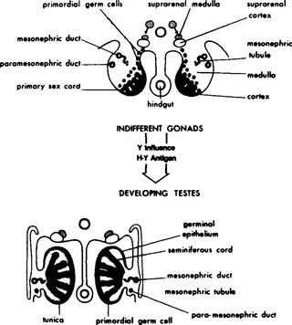
Fig. 1 Development of the biopotential gonad from the coelomic epthelium (primary sex cords), underlying mesenchymal tissue (medulla), and primordial germ cells and its differentiation to form a testes (adapted from Arey, 1965).
The primordial germ cells are visible early in the third week among the endodermal cells of the wall of the yolk sac near the origin of the allantois (Everett, 1943; Mintz and Russell, 1955, 1957). During folding of the embryo, part of the yolk sac is incorporated into the embryo and the primordial germ cells migrate along the dorsal mesentery to the gonadal ridges (Fig. 2). The germ cells are spherical and larger than the mesenchymal cells, with large vesicular nuclei and abundant cytoplasm. The germ cells multiply by mitosis during migration. In the fifth week of fetal life they begin to migrate into the underlying mesenchyme and by the end of the sixth week (13 to 15 mm) the undifferentiated or bipotential gonad is formed (Figs. 1 and 3).
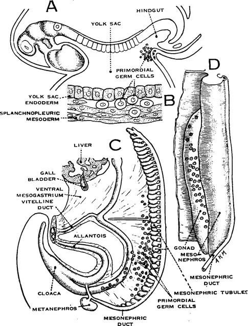
Fig. 2 Migration of primordial germ cells from the endoderm of the yolk sac (Hamilton et al., 1972).
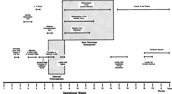
Fig. 3 Embryogenesis of the male phenotype.
Thus, the undifferentiated gonad is formed from three sources, coelomic epithelium, underlying mesenchyme, and primordial germ cells. The primordial germ cells develop into spermatogonia in the male and ova in the female; the sex cords become either seminiferous tubules or primary ovarian follicles; and the mesenchymal cells form either the Leydig cells or the theca and stromal cells in the female (Hamilton et al., 1972) (Figs. 1 and 4).
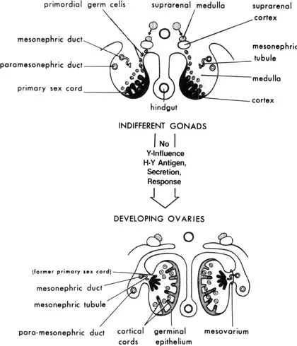
Fig. 4 The development of the bipotential gonad and its differentiation to form an ovary (adapted from Arey, 1965).
B Gonadal Differentiation
1 Testicular Differentiation
At approximately the seventh week of gestation in a 15- to 20-mm embryo, the testicular cords evolve from the primary sex cords of the indifferent gonad (Fig. 3). The Sertoli cells within each gonad enlarge, come into contact with one another, and engulf the germ cells. The distal ends of the testicular or seminiferous cords then interconnect to form a network of solid cords, the rete testes, which is in direct contact with the mesonephric tubules. At the third month of gestation the rete testes connect with the mesonephric tubules and by the sixth month the ends of the rete testes develop a lumen continuous with the mesonephric tubules which later develop into the ductuli efferen...
Table of contents
- Cover image
- Title page
- Table of Contents
- Contributors
- Editorial Board
- Copyright page
- Contributors
- Preface
- The Fetal Neuroendocrine Axis
- In Vitro Fertilization
- Factors Controlling Placental Endocrine Function in Domestic Animals
- The Fetal Thyroid
- Carbohydrate Metabolism
- Regulation of Partition of Protein During Pregnancy
- Mineral Needs of the Fetus
- Fetal Metabolism of Cortisol
- Sexual Differentiation: Normal and Abnormal
- Metabolic Errors of Adrenal Steroidogenesis
- Index
Frequently asked questions
Yes, you can cancel anytime from the Subscription tab in your account settings on the Perlego website. Your subscription will stay active until the end of your current billing period. Learn how to cancel your subscription
No, books cannot be downloaded as external files, such as PDFs, for use outside of Perlego. However, you can download books within the Perlego app for offline reading on mobile or tablet. Learn how to download books offline
Perlego offers two plans: Essential and Complete
- Essential is ideal for learners and professionals who enjoy exploring a wide range of subjects. Access the Essential Library with 800,000+ trusted titles and best-sellers across business, personal growth, and the humanities. Includes unlimited reading time and Standard Read Aloud voice.
- Complete: Perfect for advanced learners and researchers needing full, unrestricted access. Unlock 1.5M+ books across hundreds of subjects, including academic and specialized titles. The Complete Plan also includes advanced features like Premium Read Aloud and Research Assistant.
We are an online textbook subscription service, where you can get access to an entire online library for less than the price of a single book per month. With over 1.5 million books across 990+ topics, we’ve got you covered! Learn about our mission
Look out for the read-aloud symbol on your next book to see if you can listen to it. The read-aloud tool reads text aloud for you, highlighting the text as it is being read. You can pause it, speed it up and slow it down. Learn more about Read Aloud
Yes! You can use the Perlego app on both iOS and Android devices to read anytime, anywhere — even offline. Perfect for commutes or when you’re on the go.
Please note we cannot support devices running on iOS 13 and Android 7 or earlier. Learn more about using the app
Please note we cannot support devices running on iOS 13 and Android 7 or earlier. Learn more about using the app
Yes, you can access Fetal Endocrinology and Metabolism by L Martini,V. H. T. James in PDF and/or ePUB format, as well as other popular books in Medicine & Physiology. We have over 1.5 million books available in our catalogue for you to explore.