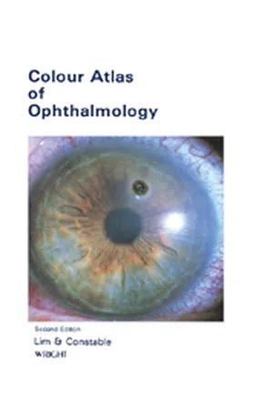
Colour Atlas of Ophthalmology
eBook - ePub

Colour Atlas of Ophthalmology

  1. 156 pages
  2. English
  3. ePUB (mobile friendly)
  4. Available on iOS & Android
eBook - ePub

Colour Atlas of Ophthalmology

Book details
Book preview
Table of contents
Citations

About This Book

Colour Atlas of Ophthalmology, Second Edition provides information pertinent to the fundamental aspects of ophthalmology. This book provides the correct diagnosis and treatment of many ocular disorders. Organized into 11 chapters, this edition begins with an overview of the process of assessment of a patient with eye disease, which includes taking a good history, examining the eyes with adequate illumination, and testing the visual function. This text then describes exophthalmos, which is the most common condition of the orbit and indicates the possibility of thyroid disease or a space-occupying lesion. Other chapters consider the common causes of ocular injuries, including injury from flying particles, sharp instruments, chemicals, and ocular injury associated with head injury. The final chapter deals with the common, therapeutic, and diagnostic ocular drugs. This book is a valuable resource for ophthalmologists, physicians, nurses, students, and all those paramedical personnel who have to deal with common eye disease.

Frequently asked questions

Simply head over to the account section in settings and click on ā€œCancel Subscriptionā€ - itā€™s as simple as that. After you cancel, your membership will stay active for the remainder of the time youā€™ve paid for. Learn more here.
At the moment all of our mobile-responsive ePub books are available to download via the app. Most of our PDFs are also available to download and we're working on making the final remaining ones downloadable now. Learn more here.
Both plans give you full access to the library and all of Perlegoā€™s features. The only differences are the price and subscription period: With the annual plan youā€™ll save around 30% compared to 12 months on the monthly plan.
We are an online textbook subscription service, where you can get access to an entire online library for less than the price of a single book per month. With over 1 million books across 1000+ topics, weā€™ve got you covered! Learn more here.
Look out for the read-aloud symbol on your next book to see if you can listen to it. The read-aloud tool reads text aloud for you, highlighting the text as it is being read. You can pause it, speed it up and slow it down. Learn more here.
Yes, you can access Colour Atlas of Ophthalmology by Arthur Lim Siew Ming,Ian J Constable in PDF and/or ePUB format, as well as other popular books in Medicine & Neurology. We have over one million books available in our catalogue for you to explore.

Information

Year
2014
ISBN
9781483281285
Edition
2
Subtopic
Neurology
1

EXAMINATION

Publisher Summary

The cause for a sudden loss of vision could be vascular in nature such as retinal vein occlusion, retinal artery occlusion, or vitreous hemorrhage. It could also be due to acute glaucoma, retinal detachment, or inflammatory conditions such as acute uveitis and optic neuritis. A gradual loss of vision is usually due to a refractive error, such as myopia or presbyopia, or degenerative conditions of which cataract is the most common. Flashes are momentary flashes of light due to the stimulation of the retina and are seen in retinal tears and detachments and also in vitreous detachment. Eye pain and headaches may be due to either ophthalmic or nonophthalmic causes. Binocular diplopia is usually due to extraocular muscle paralysis. Monocular diplopia is caused by disease in the eyeball, such as early cataract, lens dislocation, or corneal opacity. The assessment of distant and near visual acuity is important as it reflects the state of the macular function (central vision). The response of light directed at one pupil in a darkened room is known as the direct pupillary response. The extraocular muscles are examined by observing the position of the eyeballs with the patient looking straight ahead. The ophthalmoscope is used to observe abnormality in the ocular media, the optic disc, the retinal vessels, the fundal background, and the macula.

INTRODUCTION

In the assessment of a patient with eye disease, it is important to take a good history, examine the eyes with adequate illumination and test the visual function.
Recently, retinal and macular diseases have become more common as causes of severe visual loss. In these cases, a fundal examination with dilatation of the pupils in a darkened room is necessary.

HISTORY

A careful history of the patientā€™s ocular symptoms is essential. His past history and general illnesses, such as diabetes and hypertension, frequently provide useful clues.
Myopia, squint, open-angle glaucoma and dystrophic conditions have a hereditary tendency which is revealed by an inquiry into the patientā€™s family history. It is also useful to take note of allergies and of the medical therapy the patient is undergoing.

OCULAR SYMPTOMS

The more important symptoms include decreased visual acuity, floaters, ocular pain, headaches, itching, flashes, watering and double vision (diplopia).

Decreased visual acuity

Decreased visual acuity must always be investigated and the cause found. The cause for a sudden loss of vision could be vascular in nature such as retinal vein occlusion, retinal artery occlusion or vitreous haemorrhage. It could also be due to acute glaucoma, retinal detachment or inflammatory conditions such as acute uveitis and optic neuritis.
Gradual loss of vision is usually due to a refractive error such as myopia or presbyopia, or to degenerative conditions of which cataract is the most common. It could also be due to macular degeneration or chronic glaucoma.

Floaters

Another common ocular symptom which calls for further investigation is the appearance of floaters usually described by the patient as small, semi-translucent particles of varying shapes moving across the visual field with the movement of the eye. Single or double floaters of many months or years are common and usually harmless. But a sudden increase in floaters, especially when associated with lightning flashes and visual loss in patients with high myopia or in the elderly, suggests retinal disease, particularly retinal detachment.

Flashes

Flashes are momentary flashes of light due to stimulation of the retina and are seen in retinal tears and detachments and also in vitreous detachment. Other sensations of light may arise from m...

Table of contents

  1. Cover image
  2. Title page
  3. Table of Contents
  4. Copyright
  5. THE AUTHORS
  6. PREFACE
  7. PREFACE TO SECOND EDITION
  8. ACKNOWLEDGEMENT
  9. Chapter 1: EXAMINATION
  10. Chapter 2: LID, LACRIMAL APPARATUS AND ORBIT
  11. Chapter 3: CONJUNCTIVA SCLERA AND CORNEA
  12. Chapter 4: LENS AND GLAUCOMA
  13. Chapter 5: UVEAL TRACT, RETINA AND VITREOUS
  14. Chapter 6: OCULAR MANIFESTATIONS OF SYSTEMIC DISEASES
  15. Chapter 7: NEURO-OPHTHALMOLOGY
  16. Chapter 8: EYE DISEASES IN CHILDREN
  17. Chapter 9: OCULAR INJURIES
  18. Chapter 10: REFRACTIVE ERRORS
  19. Chapter 11: OPHTHALMIC DRUGS
  20. INDEX