Neuromuscular Pathology Made Easy
eBook - ePub

Neuromuscular Pathology Made Easy

  1. 240 pages
  2. English
  3. ePUB (mobile friendly)
  4. Available on iOS & Android
eBook - ePub

Neuromuscular Pathology Made Easy

Book details
Book preview
Table of contents
Citations

About This Book

The scope of Neuromuscular Pathology continuous to expand, as evidenced by the numerous multivolume and speciality texts published in recent years. This short textbook provides a complete overview of both clinical and histological aspects of common and rare neuromuscular diseases. The objective is twofold: to provide information about neuromuscular diseases in a simplified, integrated, and rapidly accessible format suited to those initially encountering the discipline, and also to provide a clear approach using simple pictures, tables and algorithms to illustrate histological features in muscle and nerve biopsy.

This volume is conveniently divided into three sections with a total of 30 chapters. The first section deals with basic principles of neuromuscular histology and physiology, processing technique, histochemistry, and laboratory management. The second and third sections deal with neuromuscular diseases that are summarized in a stepwise approach, complemented by algorithms and organized tables.

  • A simplified, integrated, and rapidly accessible format covering both common and rare neuromuscular diseases


  • Clear simple illustrations, organized tables and algorithms to aid the reader in finding an easy approach to accurate diagnosis


  • Practical tips to facilitate histopathological diagnosis.


  • Clinical scenarios discussing common neuromuscular conditions

Neurologists, neuropathologists, trainees and medical students involved in clinical neuroscience and pathology will find this guide of practical benefit in both education and practice.

Frequently asked questions

Simply head over to the account section in settings and click on “Cancel Subscription” - it’s as simple as that. After you cancel, your membership will stay active for the remainder of the time you’ve paid for. Learn more here.
At the moment all of our mobile-responsive ePub books are available to download via the app. Most of our PDFs are also available to download and we're working on making the final remaining ones downloadable now. Learn more here.
Both plans give you full access to the library and all of Perlego’s features. The only differences are the price and subscription period: With the annual plan you’ll save around 30% compared to 12 months on the monthly plan.
We are an online textbook subscription service, where you can get access to an entire online library for less than the price of a single book per month. With over 1 million books across 1000+ topics, we’ve got you covered! Learn more here.
Look out for the read-aloud symbol on your next book to see if you can listen to it. The read-aloud tool reads text aloud for you, highlighting the text as it is being read. You can pause it, speed it up and slow it down. Learn more here.
Yes, you can access Neuromuscular Pathology Made Easy by Maher Kurdi, Maher Kurdi in PDF and/or ePUB format, as well as other popular books in Medicine & Pathology. We have over one million books available in our catalogue for you to explore.

Information

Publisher
CRC Press
Year
2021
ISBN
9781000346558
Edition
1
Subtopic
Pathology

Part I

General

Chapter 1
Muscle and Nerve Histology
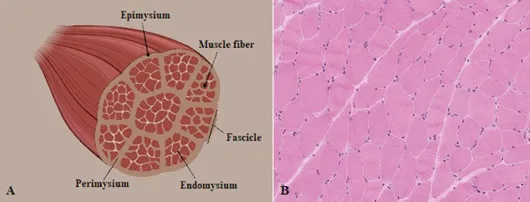
1.1Muscle Histology

Muscle tissue is differentiated into smooth, cardiac, and skeletal muscles. This differentiation embryonically starts to develop from mesoderm during the 7th week of gestation and it completely forms by the 28th week of gestation. The nuclei take their peripheral positions after 20 weeks of gestation. Skeletal muscle is elongated on longitudinal sections and striated in cross section. The entire portion is enclosed in a connective tissue sheath called epimysium, which is composed of extracellular matrix and irregular collagens. The epimysium connects into the tendon sheath and divides the muscle into groups of bundles, or fascicles, separated from each other by perimysium (Figure 1.1). The myotendinous junction area sometimes mimics dystrophy (pseudomyopathy), as it may show alternative features of hypertrophic and atrophic fibers, excess fibrous tissue, and multiple internal nuclei (Figure 1.2).
Figure 1.1
Figure 1.1(a) An illustrated photograph of skeletal muscle layers. (b) Cross section of normal skeletal muscle shows the muscle fibers within the fascicles. (H&E ×20.)
Figure 1.2
Figure 1.2Myotendinous junction with hypertrophic and atrophic fibers, multiple internal nuclei, and increased connective tissue. (H&E ×40.)
Each group of muscle fascicles is composed of uniform muscle fibers called myocytes. They are separated from each other by a network of reticular fibers and extracellular matrix, called endomysium.
Each muscle fiber contains either a single nucleus or multiple nuclei and a cytoplasm called sarcoplasm, and it is surrounded by a plasma membrane called sarcolemma. The nuclei are usually peripherally located, heterochromatic, and containing fine nucleoli and stippled nucleoplasm. They stain blue with hematoxylin and red with Gomori trichrome. Centrally positioned nuclei for more than 3% of a whole fascicle is considered abnormal.
The sarcoplasm contains multiple organelles including mitochondria, sarcoplasmic reticulum, Golgi apparatus, microtubules, glycogens, ribosomes, lipid droplets, and myofibrils. The mitochondria are located at the level of the I-band. They present in type I fibers more than type II fibers, but this is not considered a consistent distinguishing feature.
The sarcolemma consists of an inner plasma membrane (plasmalemma) and outer membrane (external basal lamina). The plasmalemma is an excitable membrane composed of a lipid bilayer and a variety of ion channels and proteins present on the cytoskeleton. Some of these sarcolemmal proteins are summarized in Table. 1.1 and Figure 1.3.
Table 1.1 Intracellular and extracellular sarcolemmal proteins associated with each muscle fiber.
Protein
Structure
Interaction site
Dystrophin
Sarcolemmal protein with four domains (actin-binding, central rod, carboxyl terminus, cysteine-rich domain)
Sarcoglycan, dystroglycan
syntrophin, dystrobrevin
Sarcoglycan
Transmembrane protein that has four subtypes: α, β, γ, δ
Dystrophin, filamin-C
Dystroglycan
Transmembrane protein (α, β). α-II laminin (merosin)
Dystrophin, caveolin-3
Dysferlin
Type II transmembrane protein
Caveolin-3
Caveolin-3
Protein found in caveolae
Dysferlin, RYR1
Myotilin
Sarcomeric Z-disc protein
Filamin-C, α-actinin
Emerin
Nuclear membrane protein anchored to cytoskeleton
Actin, lamin A/C, CTNNB1
Abbreviations: RYR1: ryanodine receptor 1; CTNNB1: catenin beta-1 protein.
Note: The interaction site is the binding site where each protein interacts with another one.
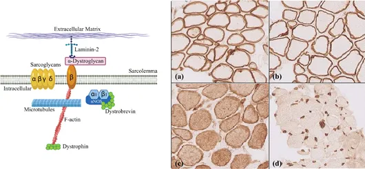
Figure 1.3
Figure 1.3(Left) A diagram of sarcolemmal muscle proteins. (Right) Groups of important muscle proteins: (a) dystrophin; (b) sarcoglycan; (c) dysferlin; (d) emerin (×40). This figure was created with Biorender.com.
The plasmalemma extends deeps into the muscle fibers, forming T-tubules that carry the depolarization of action potential inside the fibers. Hence, a T-tubule with two terminal cisterns forms a triad. The triad has a voltage-gated calcium channel and ryanodine receptor. T-tubules and sarcoplasmic reticulum are essential components involved in muscle contractio...

Table of contents

  1. Cover
  2. Half Title
  3. Title Page
  4. Copyright Page
  5. Contents
  6. List of Contributors
  7. Preface
  8. Acknowledgments
  9. Abbreviations
  10. Part I General
  11. Part II Muscle
  12. Part III Nerve
  13. Index