![]()
Part One
![]()
Chapter 1
Introduction to the Nervous System
Let’s begin by examining the divisions of the nervous system.
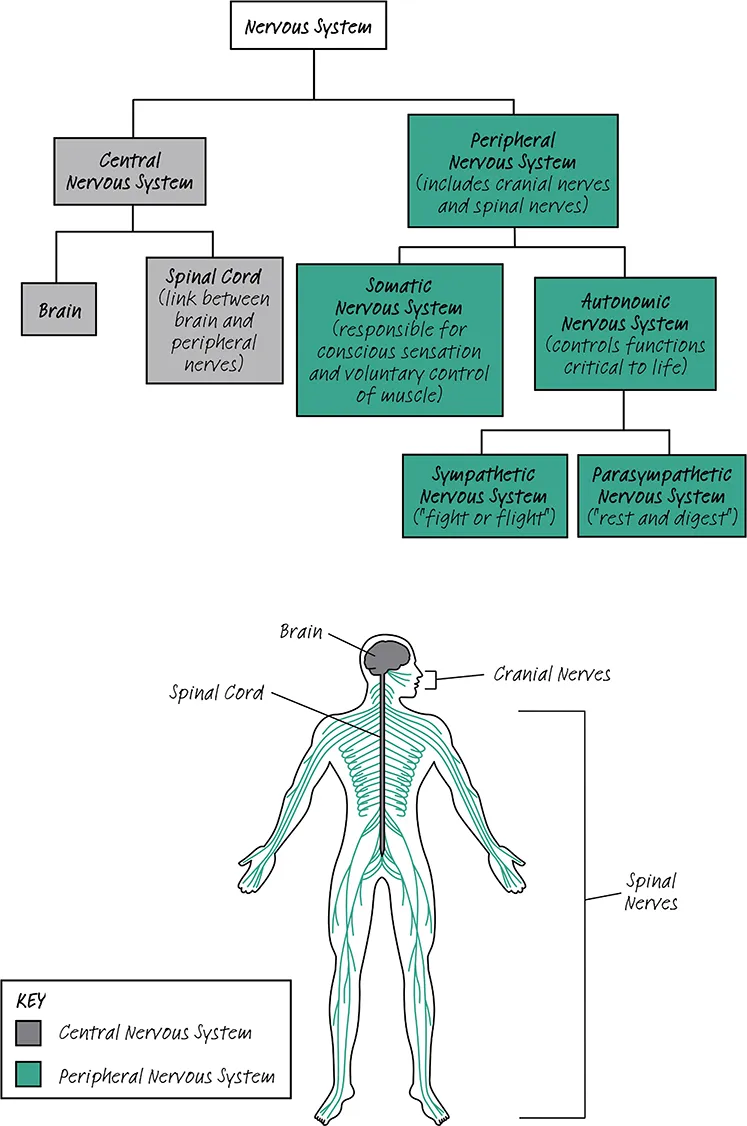
The human nervous system is first divided into the central nervous system (CNS) and the peripheral nervous system (PNS). The CNS is made up of the brain and spinal cord. The PNS is made up of cranial nerves (CN), which innervate the head, and spinal nerves, which innervate the body. The function of the PNS is to transmit information to and from the CNS. Since many nerves feed into the spinal cord, it is sometimes called the “bridge” between the PNS and CNS.
The PNS can be further subdivided into the autonomic nervous system, and the somatic nervous system. The autonomic nervous system is not under conscious control; it automatically controls all of the functions needed to sustain life (i.e., blood pressure, respiratory rate, digestion and excretion). The autonomic nervous system can be further subdivided into the sympathetic nervous system, which readies you for an impeding conflict, and the parasympathetic nervous system, which conserves energy and regulates growth.
The somatic nervous system, by contrast, is under conscious control. It is responsible for conscious sensation (such as feeling something is cold, or that something is sharp) as opposed to the unconscious sensation of the autonomic nervous system (such as your carotid bulbs sensing decreased blood perfusion to the brain). It is also responsible for the voluntary control of muscles. The autonomic and somatic systems act through the CN in the head as well as the spinal nerves in the body.
The autonomic system and the somatic system should be thought of as distinct entities. However, they can both travel together in the same nerve. For example, CN III is considered to have a somatic and autonomic component. However, this is usually for just a short part of the nerve pathway, and the components soon separate. We will examine this further in Chapter 3.
The CNS is made up of the brain and spinal cord.
The PNS is composed of the somatic nervous system, under conscious control, and the autonomic nervous system, which is not under conscious control.
The somatic system and the autonomic system act both through cranial nerves and spinal nerves.
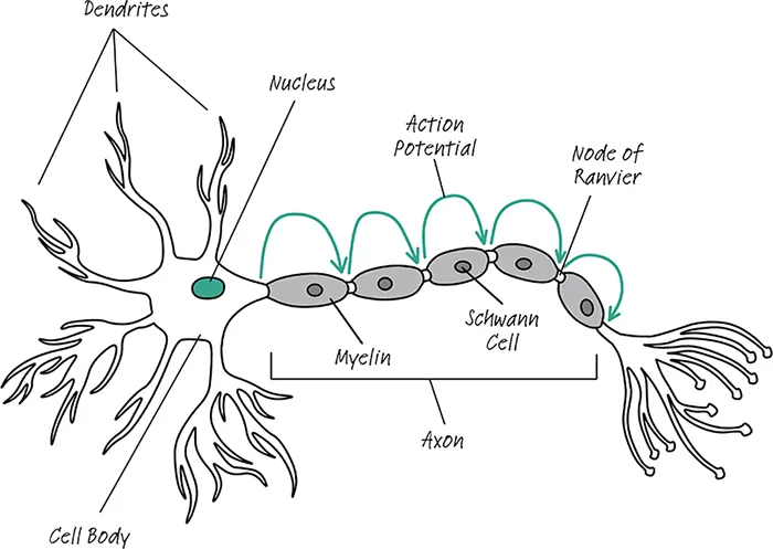
Before we continue, it is useful to go over the anatomy of a neuron. A neuron is a single cell composed of three main parts – dendrites, a single cell body and a single axon. Dendrites transmit information signals toward the cell body of the neuron. Axons transmit information away from the cell body, in the form of an electrical impulse called an action potential. Action potentials are caused by transient changes in the voltage measured between a neuron’s cell membrane and the exterior environment. The cell body contains the nucleus and all the cellular machinery necessary to sustain the neuron.
An axon is covered in myelin, which is produced in the PNS by the Schwann cell. In the CNS, myelin is produced by another kind of cell, the oligodendrocyte. Nodes of Ranvier are small areas along the axon that do not contain myelin. Myelin allows for much more rapid transmission of action potentials, as they can jump between Nodes of Ranvier as shown in Fig. 1.2.
Many dendrites can feed into one cell body, but each cell body has only one axon. Even though most books draw the dendrites as being quite short, they can be very long, even longer than the axon. It is important to note that one neuron spans the entire length of any individual nerve. Thus, the neurons of the sciatic nerve are over 1 meter (over 3 feet) long!
The nomenclature of neuron cell bodies is important and can be a cause of confusion. In the CNS a collection of cell bodies is called a nucleus. In the PNS a collection of cell bodies is called a ganglion (pl. ganglia). This difference is illustrated nicely by the autonomic nervous system, as we will see in a few pages.
Dendrites transmit information towards the cell body and axons transmit information away from the cell body.
Axons are covered in myelin, which is made by Schwann cells in the PNS and oligodendrocytes in the CNS.
A collection of cell bodies is called a nucleus in the CNS and a ganglion in the PNS.
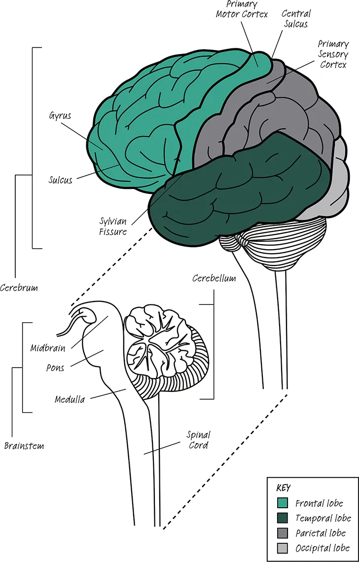
As we saw earlier, the CNS is composed of the brain and spinal cord. The brain itself is made up of the cerebrum, brainstem and cerebellum.
The cerebrum is what most people picture when they think of the brain. It is divided into two identical hemispheres: one on the left side and one on the right side. The outer layer of the cerebrum is called the cortex and made up of folded layers of tissue called gray matter. Gray matter houses neuronal cell bodies and initiates neuronal impulses. The other type of tissue in the CNS is white matter, which contains the axons that transmit the impulses initiated from the gray matter.
If you examine the cerebrum shown in Fig. 1.3, you will see it is composed of many ridges and valleys. A ridge is called a gyrus and a valley is called a sulcus. The central sulcus is important because the gyrus anterior to it is the primary motor cortex, which initiates all motor functions. The gyrus posterior to the central sulcus is the primary sensory cortex, which receives all sensory input.
The cerebrum is divided into lobes by various anatomical landmarks. The frontal lobe extends back to the central sulcus, and inferiorly until the Sylvian fissure. The frontal lobe is responsible for thinking, planning and all motor control. The parietal lobe is posterior to the central sulcus but doesn’t have a specific defining landmark between it and the temporal or occipital lobes. The parietal lobe receives and integrates all sensory information. The temporal lobe sits inferior to the Sylvian fissure, but doesn’t have a defined posterior border; it creates and stores new memory and is involved in emotional regulation. It also contributes to the understanding of language. Finally, the occipital lobe serves as the visual processing center.
The brainstem sits just inferior to the cerebrum, and is divided into three sections called the midbrain, pons and medulla. The brainstem regulates all life preserving functions and controls consciousness. It also provides all innervation to the head via the CN. Just inferior to it sits the cerebellum, which is responsible for coordinating smooth, voluntary movement.
Just as the brainstem provides all innervation to the head via the CN, the spinal cord provides all innervation to the body via the spinal nerves.
The brain is made up of the cerebrum, brainstem and cerebellum.
The cerebrum is divided into four lobes: frontal, parietal, temporal and occipital.
The brainstem is divided into the midbrain, pons and medulla.
The brainstem provides all innervation to the head, and the spinal cord provides all innervation to the body.
The PNS consists of the CN and the spinal nerves. We will discuss the CN in detail in Chapter 3. In order to understand the spinal nerves, we need to briefly discuss their relation to the spinal cord.
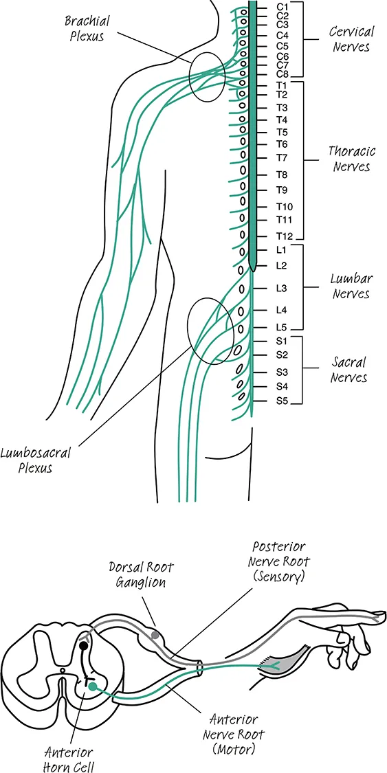
A total of 31 paired spinal nerves exit the spinal cord. Because of morphological differences that occur in the spinal cord, the spinal cord and its associated nerves are divided into four different sections: cervical, thoracic, lumbar and sacral. There are 8 cervical nerves, 12 thoracic nerves, 5 lumbar nerves and 5 sacral nerves (in addition there is one coccygeal nerve, but it serves no clinically relevant purpose so it is usually ignored). Nerves are named by stating which group they belong to, and the relative position of the nerve in the group; for example the 6th nerve of the cervical group is called Cervical 6 (abbreviated C6).
If we take a closer look at the spinal cord we will see that a total of four nerve roots exit the spinal cord (two on each side). There is an anterior nerve root, which carries motor signals to muscle, and a posterior nerve root, which carries sensory information to the brain. The nucleus for the motor neuron lies in a special area of the gray matter, called the a...