This book focuses on the impact of nutritional disorders on the nervous system. Nutritional disorders are caused due to poverty, famine, infestations, ignorance in the developing world and due to food faddism, isolation, depression, addictions, and comorbidities in the developed countries. This book has chapters on various disorders covering basic knowledge, their clinical manifestations, basis and etiology, laboratory diagnosis, method of treatment and prognosis. It provides the guidelines to students and clinicians for dealing with such disorders which are easily preventable and amenable to treatment whose early diagnosis and management can avert morbidity and mortality.
Key Features
Deals with the unexplored topic of the neurological impact of nutritional disorders
Will be essential for neurologists, general physicians, and pediatricians
Includes key illustrated examples from authors' clinical practice.
Trusted by 375,005 students
Access to over 1.5 million titles for a fair monthly price.
Role of nutrition in the growth and development of nervous system
Introduction
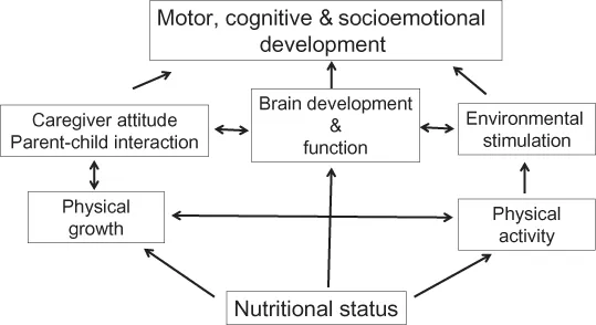
Nutrition is essential throughout life, and is especially important during pregnancy, infancy and early childhood for the development of nervous system. Development of brain is related to the development of cognitive and motor functions, and socioemotional skills during childhood and adulthood. Nutritional deficiencies during pregnancy and infancy can affect cognitive and behavioral functions, which predict school performance as well as performance in later life. This chapter highlights the importance of nutrition in different stages of the nervous system development.
Structural development
The biological time scale of human development requires observation in a large number of individuals. Since it is difficult to accurately decide the time of conception, the gestational age (GA) may not be precise. The average human GA is 40 weeks. In early prenatal life, the chronological and biological scales have special importance. The development in this period is so fast that there is change every day. During infancy, the tempo of development is slower than that in the parental life, but it is faster than that in childhood.
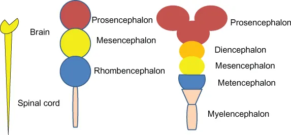
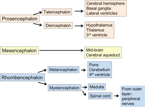
Central nervous system is developed as a specialized ectoderm, called “neuroectoderm”. The thickening of neuroectoderm results in the formation of neural plates, which in turn fuse to form a neural tube. The cephalic portion of the neural tube enlarges to form the brain, and the caudal portion narrows to form the spinal cord and peripheral nerves. There are three dilatations in the cranial part: prosencephalon, mesencephalon and rhombencephalon. The prosencephalon differentiates into telencephalon and diencephalon. The telencephalon differentiates into cerebral hemispheres, corpus striatum and cerebral ventricles. The structures develop from the diencephalon are thalamus, hypothalamus and related structures, including the third ventricle. The mesencephalon gives rise to midbrain and cerebral aqueduct. The rhombencephalon has two parts: metencephalon (from which pons, cerebellum and the fourth ventricle develop) and the myelencephalon (which differentiates into medulla and spinal cord) (Figures 1.1 and 1.2). The outer layer of the spinal cord develops into peripheral nerves. The lateral aspects of neural crest and adjacent cells differentiate into dorsal root ganglia, adrenal medulla, sympathetic and parasympathetic ganglia, Schwann cells and neurolemmal sheets. The neural tube with single-cell lining develops into three layers: ependymal matrix, mental and meningeal layers. The neurons are derived from the mental layer.
Figure 1.1 Schematic diagram shows the development of different parts of notochord.
Figure 1.2 Origin of different structures of the nervous system from prosencephalon, mesencephalon and rhombencephalon.
The development of nervous system is a continuous process rather than a stepwise event. Several functions develop in parallel, and any dissociation is abnormal.
Embryonal and fetal development
At 3 weeks of embryonic life, there are neuroblastic differentiation, migration and neuronal multiplication, which have a genetic basis. The neuronal proliferation occurs at an average speed of 250,000/minute for several days to weeks. The neurons become bipolar and migrate to the marginal layer through radial glial cells to form the cerebral cortex. Migration is completed by the fifth fetal month when the cortex contains billions of postmitotic cells. During the mid-fetal life, cerebral hemispheres develop into a deeply sulcated structure. The major sulci such as Sylvian, Rolandic and calcarine sulci achieve an adult configuration by the fifth month. Secondary sulci develop by 6–7 months of fetal life, and tertiary sulci also develop by 8–9 months. Simultaneously, there is an organization of cortical and neuronal masses. Synaptogenesis and axonal path finding take place subsequently by 30th week. The cytoarchitectural pattern of the cerebral cortex develops and differentiates neurons of the cortical region, e.g., primary motor, sensory and auditory cortices.
The time scale of myelin development is not as clearly known as that of the neuronal development. By 10th week, spinal roots and nerves are myelinated, which correspond with the reflex motor activity. Subsequently, segmental and intersegmental fibers are myelinated followed by the myelination of ascending and descending tracts that start from the brainstem. By 28th–30th week, vestibulo-cochlear pathways are myelinated; by 37th week, myelination of spinocerebellar and dentatorubral tracts takes place. By 40th week of gestation, the visual system starts myelinating, which is completed by 3 months of postnatal life. The corticospinal tracts are myelinated by 18 months of postnatal life. Posterior frontal and parietal lobes are myelinated by 40th week of gestation, which is followed by the myelination of other parts of the brain. By 2 years, the myelination of the nervous system is complete. The myelination of intra- and interhemispheric fibers continues till 10 years of age or beyond. The density of neurons increases up to 15 months of postnatal life; thereafter, it decreases with increasing dendritic arborization and synaptogenesis. The growth of human embryo and fetus is presented in Table 1.1. The neurons begin to function before myelination. By the age of 12–15 years, the weight of the brain in females is 1230–1275 g and in males 1350–1410 g. The development of brain structure at different time points is shown in Figure 1.3.
Table 1.1 Growth of nervous system in human embryo and fetus
Fetal age (days)
Length (mm)
Nervous system development
18
1.5
Neural groove and tube
21
3
Optic vesicles
26
3
Anterior neuropore closure
27
3.3
Posterior neuropore closure
31
4.3
Anterior horn cell appears
Anterior and posterior roots
35
5
Five vesicles
42
13
Primordium of cerebellum
56
25
Differentiation of cortex and meninges
150
225
Primary cerebral fissures
180
230
Cerebral myelination
8–9 months
240
Growth of brain and myelination
Figure 1.3 Organization of cellular and functional structures at different natal and postnatal periods of life.
Requirement of nutrients in the developing brain
Growth factors and nutrients play an important role in the development of brain during fetal and early postnatal life. Some nutrients play a greater role, such as protein, carbohydrate, fat, iron, zinc, copper, iodine, selenium, choline, vitamin A, folate and cobalamin. The effects of nutritional deficiency may depend on timing, dose and duration of deficiency. There are three stages of neuronal involvement in malnutrition:
Stage 1: Structure of cell and process may be intact but its function may be impaired.
Stage 2: Demyelination with an intact axis cylinder.
Stage 3: Demyelination, destruction of axis cylinder and cell death.
Replenishment of nutrients in stage 1 rapidly restores the function. In stages 2 and 3, there may be a permanent deficit. During 24–42 weeks of GA, brain is particularly vulnerable to nutritional deficiency, because myelination and synaptogenesis take place during this period. During the late fetal and early neonatal life, hippocampus, visual, auditory cortex and corpus striatum have a fast pace of development with regard to morphogenesis and synaptogenesis. Hippocampus is the first region to have a cortico-cortical connectivity and becomes functional by 1 month of postnatal life. Myelination may also be affected if there is a nutritional deprivation. Nutrients at a particular concentration may be beneficial at one time, but can as well be toxic at the other time. Iron and certain other nutrients are regulated in a narrow range, highlighting...
Table of contents
Cover
Half Title
Title Page
Copyright Page
Dedication
Table of Contents
Preface
Acknowledgments
About the authors
1 Role of nutrition in the growth and development of nervous system
2 Protein energy malnutrition
3 Thiamine deficiency neurological disorders
4 Niacin deficiency: Pellagra
5 Neurological manifestations of pyridoxine deficiency
6 Neurological effects of Vitamin B12 deficiency
7 Vitamin D deficiency
8 Vitamin E deficiency and its neurological effects
9 Neurological manifestations of fluorosis
10 Iodine deficiency disorders and their neurological consequences
11 Neurological consequences of copper and zinc deficiency
12 Water as a nutrient in health and disease
13 Electrolytes in health and disease
14 Specific dietary management of certain neurological disorders
Index
Frequently asked questions
Yes, you can cancel anytime from the Subscription tab in your account settings on the Perlego website. Your subscription will stay active until the end of your current billing period. Learn how to cancel your subscription
No, books cannot be downloaded as external files, such as PDFs, for use outside of Perlego. However, you can download books within the Perlego app for offline reading on mobile or tablet. Learn how to download books offline
Perlego offers two plans: Essential and Complete
Essential is ideal for learners and professionals who enjoy exploring a wide range of subjects. Access the Essential Library with 800,000+ trusted titles and best-sellers across business, personal growth, and the humanities. Includes unlimited reading time and Standard Read Aloud voice.
Complete: Perfect for advanced learners and researchers needing full, unrestricted access. Unlock 1.5M+ books across hundreds of subjects, including academic and specialized titles. The Complete Plan also includes advanced features like Premium Read Aloud and Research Assistant.
Both plans are available with monthly, semester, or annual billing cycles.
We are an online textbook subscription service, where you can get access to an entire online library for less than the price of a single book per month. With over 1.5 million books across 990+ topics, we’ve got you covered! Learn about our mission
Look out for the read-aloud symbol on your next book to see if you can listen to it. The read-aloud tool reads text aloud for you, highlighting the text as it is being read. You can pause it, speed it up and slow it down. Learn more about Read Aloud
Yes! You can use the Perlego app on both iOS and Android devices to read anytime, anywhere — even offline. Perfect for commutes or when you’re on the go. Please note we cannot support devices running on iOS 13 and Android 7 or earlier. Learn more about using the app
Yes, you can access Neurological Consequences of Nutritional Disorders by U. K. Misra,J. Kalita in PDF and/or ePUB format, as well as other popular books in Medicine & Nutrition, Dietics & Bariatrics. We have over 1.5 million books available in our catalogue for you to explore.