Aromadermatology
eBook - ePub

Aromadermatology

Aromatherapy in the Treatment and Care of Common Skin Conditions

  1. 145 pages
  2. English
  3. ePUB (mobile friendly)
  4. Available on iOS & Android
eBook - ePub

Aromadermatology

Aromatherapy in the Treatment and Care of Common Skin Conditions

Book details
Book preview
Table of contents
Citations

About This Book

This comprehensive, evidence-based guide promotes an integrative approach to using complementary therapies with conventional medicines.

It increases awareness of the sound scientific basis to aromatherapy with a wealth of data, and contains practical information for treatment.

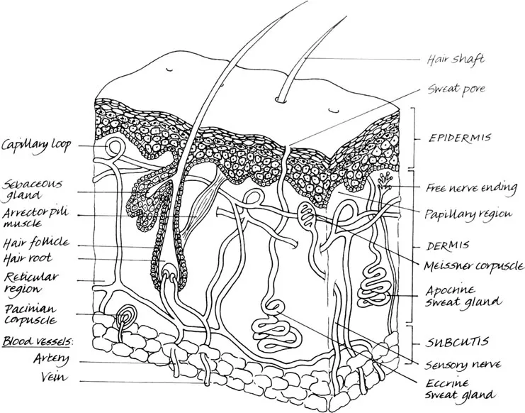
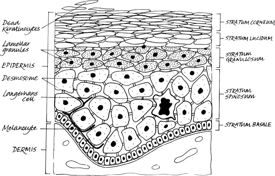
Contents include: Skin structure and function * Essential oil sciences in context * Aromadermatology and safety issues * The essentials of aromatic formulations * Skin-care essentials * Skin and the psyche * Skin infections * Childhood skin complaints * Inflammatory disorders * Wound care * Nails, hair and sebaceous glands

'With the growing interest in aromatherapy, it is important that therapists and healthcare professionals are able to offer a valid rationale when integrating essential oils into clinical care. Sound knowledge of bio-chemical principles and the ability to critically appraise and apply relevant research are fundamental requirements. This book offers a comprehensive, in-depth view of current knowledge. The authors have skilfully woven research and clinical application. A range of therapeutic possibilities is explored and offers practitioners alternative approaches to the management of skin conditions. These include detailed discussions on different methods of application. I hope that this book will become a standard text on both pre-qualifying and CPD courses in aromatherapy.' – Angela Avis, in her Foreword

'This well-illustrated, thorough and authoritative text is written in a language and style that is clear and accessible to a variety of healthcare practitioners. A thorough understanding of dermatology underpins the book, and both current research and clinical knowledge are elegantly applied to the skin conditions discussed.' – Robert Tisserand, in his Foreword

Frequently asked questions

Simply head over to the account section in settings and click on “Cancel Subscription” - it’s as simple as that. After you cancel, your membership will stay active for the remainder of the time you’ve paid for. Learn more here.
At the moment all of our mobile-responsive ePub books are available to download via the app. Most of our PDFs are also available to download and we're working on making the final remaining ones downloadable now. Learn more here.
Both plans give you full access to the library and all of Perlego’s features. The only differences are the price and subscription period: With the annual plan you’ll save around 30% compared to 12 months on the monthly plan.
We are an online textbook subscription service, where you can get access to an entire online library for less than the price of a single book per month. With over 1 million books across 1000+ topics, we’ve got you covered! Learn more here.
Look out for the read-aloud symbol on your next book to see if you can listen to it. The read-aloud tool reads text aloud for you, highlighting the text as it is being read. You can pause it, speed it up and slow it down. Learn more here.
Yes, you can access Aromadermatology by Janetta Bensouilah, Philippa Buck in PDF and/or ePUB format, as well as other popular books in Medicina & Medicina alternativa y complementaria. We have over one million books available in our catalogue for you to explore.

Information

Publisher
Routledge
Year
2021
ISBN
9781000477085

Chapter 1

Skin structure and function

Introduction

The integument or skin is the largest organ of the body, making up 16% of body weight, with a surface area of 1.8 m2. It has several functions, the most important being to form a physical barrier to the environment, allowing and limiting the inward and outward passage of water, electrolytes and various substances while providing protection against micro-organisms, ultraviolet radiation, toxic agents and mechanical insults. There are three structural layers to the skin: the epidermis, the dermis and subcutis. Hair, nails, sebaceous, sweat and apocrine glands are regarded as derivatives of skin (see Figure 1.1). Skin is a dynamic organ in a constant state of change, as cells of the outer layers are continuously shed and replaced by inner cells moving up to the surface. Although structurally consistent throughout the body, skin varies in thickness according to anatomical site and age of the individual.
Image
Figure 1.1 Cross-section of the skin.
Table 1.1 Layers of the skin.
Skin layer
Description
Epidermis
The external layer mainly composed of layers of keratinocytes but also containing melanocytes, Langerhans cells and Merkel cells.
Basement membrane
The multilayered structure forming the dermoepidermal junction.
Dermis
The area of supportive connective tissue between the epidermis and the underlying subcutis: contains sweat glands, hair roots, nervous cells and fibres, blood and lymph vessels.
Subcutis
The layer of loose connective tissue and fat beneath the dermis.

Skin anatomy

The epidermis is the outer layer, serving as the physical and chemical barrier between the interior body and exterior environment; the dermis is the deeper layer providing the structural support of the skin, below which is a loose connective tissue layer, the subcutis or hypodermis which is an important depot of fat (see Table 1.1).

Epidermis

The epidermis is stratified squamous epithelium. The main cells of the epidermis are the keratinocytes, which synthesise the protein keratin. Protein bridges called desmosomes connect the keratinocytes, which are in a constant state of transition from the deeper layers to the superficial (see Figure 1.2). The four separate layers of the epidermis are formed by the differing stages of keratin maturation. The epidermis varies in thickness from 0.05 mm on the eyelids to 0.8–1.5 mm on the soles of the feet and palms of the hand. Moving from the lower layers upwards to the surface, the four layers of the epidermis are:
  • stratum basale (basal or germinativum cell layer)
  • stratum spinosum (spinous or prickle cell layer)
  • stratum granulosum (granular cell layer)
  • stratum corneum (horny layer).
In addition, the stratum lucidum is a thin layer of translucent cells seen in thick epidermis. It represents a transition from the stratum granulosum and stratum corneum and is not usually seen in thin epidermis. Together, the stratum spinosum and stratum granulosum are sometimes referred to as the Malphigian layer.
Image
Figure 1.2 Layers of the epidermis.

Stratum basale

The innermost layer of the epidermis which lies adjacent to the dermis comprises mainly dividing and non-dividing keratinocytes, which are attached to the basement membrane by hemidesmosomes. As keratinocytes divide and differentiate, they move from this deeper layer to the surface. Making up a small proportion of the basal cell population is the pigment (melanin) producing melanocytes. These cells are characterised by dendritric processes, which stretch between relatively large numbers of neighbouring keratinocytes. Melanin accumulates in melanosomes that are transferred to the adjacent keratinocytes where they remain as granules. Melanin pigment provides protection against ultraviolet (UV) radiation; chronic exposure to light increases the ratio of melanocytes to keratinocytes, so more are found in facial skin compared to the lower back and a greater number on the outer arm compared to the inner arm. The number of melanocytes is the same in equivalent body sites in white and black skin but the distribution and rate of production of melanin is different. Intrinsic ageing diminishes the melanocyte population.
Merkel cells are also found in the basal layer with large numbers in touch-sensitive sites such as the fingertips and lips. They are closely associated with cutaneous nerves and seem to be involved in light touch sensation.

Stratum spinosum

As basal cells reproduce and mature, they move towards the outer layer of skin, initially forming the stratum spinosum. Intercellular bridges, the desmosomes, which appear as ‘prickles’ at a microscopic level, connect the cells. Langerhans cells are dendritic, immunologically active cells derived from the bone marrow, and are found on all epidermal surfaces but are mainly located in the middle of this layer. They play a significant role in immune reactions of the skin, acting as antigen-presenting cells.

Stratum granulosum

Continuing their transition to the surface the cells continue to flatten, lose their nuclei and their cytoplasm appears granular at this level.

Stratum corneum

The final outcome of keratinocyte maturation is found in the stratum corneum, which is made up of layers of hexagonal-shaped, non-viable cornified cells known as corneocytes. In most areas of the skin, there are 10–30 layers of stacked corneocytes with the palms and soles having the most. Each corneocyte is surrounded by a protein envelope and is filled with water-retaining keratin proteins. The cellular shape and orientation of the keratin proteins add strength to the stratum corneum. Surrounding the cells in the extracellular space are stacked layers of lipid bilayers (see Figure 1.3).
The resulting structure provides the natural physical and water-retaining barrier of the skin. The corneocyte layer can absorb three times its weight in water but if its water content drops below 10% it no longer remains pliable and cracks. The movement of epidermal cells to this layer usually takes about 28 days and is known as the epidermal transit time.

Dermoepidermal junction/basement membrane

This is a complex structure composed of two layers. Abnormalities here result in the expression of rare skin diseases such as bullous pemphigoid and epidermolysis bullosa. The structure is highly irregular, with dermal papillae from the papillary dermis projecting perpendicular to the skin surface. It is via diffusion at this junction that the epidermis obtains nutrients and disposes of waste. The dermoepidermal junction flattens during ageing which accounts in part for some of the visual signs of ageing.

Dermis

The dermis varies in thickness, ranging from 0.6 mm on the eyelids to 3 mm on the back, palms and soles. It is found below the epidermis and is composed of a tough, supportive cell matrix. Two layers comprise the dermis:
  • a thin papillary layer
  • a thicker reticular layer.
The papillary dermis lies below and connects with the epidermis. It contains thin loosely arranged collagen fibres. Thicker bundles of collagen run parallel to the skin surface in the deeper reticular layer...

Table of contents

  1. Cover
  2. Title Page
  3. Copyright Page
  4. Table of Contents
  5. Forewords
  6. Preface
  7. About the authors
  8. Acknowledgements
  9. Botanical terminology
  10. List of abbreviations
  11. 1 Skin structure and function
  12. 2 Essential oil sciences in context
  13. 3 Aromadermatology and safety issues
  14. 4 The essentials of aromatic formulations
  15. 5 Skin-care essentials
  16. 6 Skin and the psyche
  17. 7 Skin infections
  18. 8 Childhood skin complaints
  19. 9 Inflammatory disorders
  20. 10 Wound care
  21. 11 Nails, hair and sebaceous glands
  22. Glossary
  23. Appendix
  24. Some voluntary organisations and support groups
  25. Index