Contemporary Scleral Lenses: Theory and Application, provides comprehensive information about scleral lenses. Chapters of this volume have been contributed by renowned scleral lens experts and cover a variety of interesting topics. These topics include the history and evolution of scleral lenses, basic scleral lens structure, optics and customizable features of scleral lenses, analysis of ocular surface shape, ocular surface topography and advances in optometry technology. These topics give readers an explanation of how to utilize diagnostic equipment in optometry practice and enables practitioners to employ a scientific and objective approach to scleral lens fitting.
Key features of this volume include:
- A straightforward approach to ophthalmic examination flow, evaluation and documentation
- A review of Scleral lens care and handling
- Descriptions of a variety of complex medical and ocular indications for scleral lenses
- Strategic tips to promote your own scleral lens practice
- A unique perspective of esteemed corneal specialists regarding the collaborative care of the patient
This textbook is a suitable reference for ophthalmology students and practitioners. This text will assist practitioners in enhancing their scleral lens practice by providing them useful information for improving patient vision, ocular surface rehabilitation and quality of life.
eBook - ePub
Contemporary Scleral Lenses: Theory and Application
- English
- ePUB (mobile friendly)
- Available on iOS & Android
eBook - ePub
Contemporary Scleral Lenses: Theory and Application
About this book
Trusted by 375,005 students
Access to over 1.5 million titles for a fair monthly price.
Study more efficiently using our study tools.
Information
Topic
MedicineSubtopic
Opthalmology & OptometryHistory of Scleral Lenses
Timothy J. Bowden1, †, Melissa Barnett2, *
1 Bowden & Lowe, Kent, UK
2 University of California, Davis, Sacramento, CA, USA
Abstract
Scleral lenses have existed for centuries, longer than any other type of contact lens. They were first conceptualized by Leonardo DaVinci in the early 16th century and were first manufactured in Europe in the late 1800s. The first scleral lenses were blown glass scleral shells without power made in 1887 by Fredrich A. Müller and Albert C. Müller. The primary indication for these scleral lenses was to manage ocular surface disease. In 1889, Adolf Eugen Gaston Fick described the use of scleral lenses with optics added to correct vision. In 1889, Eugene Kalt described contact lenses as orthopedic appliances for the treatment of keratoconus. In that same year, August Müeller created a scleral lens for himself to correct his own 14D of high myopia. Modern advances of scleral lenses have overcome their previous shortcomings, including lens-induced corneal edema due to poor transmissibility of oxygen through the lens and poor reproducibility. Contemporary scleral lenses have re-emerged from a long history of contact lens successes and some failures. In recent years, there has been a burst of new designs and innovations worldwide. With modern materials, manufacturing, and advanced scanning equipment, scleral lenses are now very innovative and a million miles away from their early beginnings.
Keywords: Air bubble, Artificial eye, Blown glass contact lens, Cast, Clearance, Experiment, Fitting, Gas permeable, Glass, Glass mask, Impression, Keratoconus, Lens making, Material, Mold, Optician, PMMA, Refractive error, Scleral shell, Transitions.
* Corresponding author Melissa Barnett: University of California, Davis, Sacramento, CA, USA; Tel: 916-734- 4641; Fax: 916-565-1640; E-mail: [email protected]
† Deceased
INTRODUCTION
A question that is often asked is: when and by whom were contact lenses invented? Well, contact lenses were not so much invented as evolved. There have been many stages in this development, some theoretical, but in the main much more practical. Today, contact lenses are largely used for cosmetic reasons, but they originally were designed to deal with medical problems including keratoconus and Symblepharon. Most of the early contact lenses were made by
artificial eye makers. Who can be better to make something to fit onto the front surface of an eye and under the eyelids than a person who would already have the skills required?
Why were the first contact lenses designed as scleral lenses? This was a result of them being made by artificial eye makers. The first contact lenses were supported by the sclera, and the optic portion vaulted over the cornea. They could as easily have been made as corneal lenses. In fact, this happened in Japan when Kyoichi Tanaka designed his first contact lens. He had never seen a contact lens before, so what he designed was actually a corneal lens. This was the start of what was to become Menicon. It has now turned full circle. With new materials and new measuring, fitting, and manufacturing techniques, many eye care practitioners are turning to scleral lenses for improved vision and comfort for their visually compromised patients.
Artificial eye making originated in Egypt around 2000 BC to ensure that Egyptians could see in the afterlife, and it gradually spread throughout Europe, first through Venice and later through Germany, the UK and then the US (Fig. 1). Artificial eye making was big business. Eye damage and eye loss were very common due to frequent, severe eye infections resulting from poor hygiene and no antibiotics, emerging industrial processes using lime and hot metals, and the non-availability of protective eyewear.
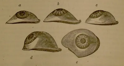
Images of artificial eyes.
“The Optician” of 28th July 1898 reported that 2,000,000 artificial eyes were produced in the German Empire each year, and a single French firm was producing 300,000 per year. “The Optician” of 1st June 1899 reported a horse being fitted with an artificial eye made of vulcanite: “There is nothing a lover of horses dislikes more than a disfigured animal”. It is no co-incidence that the earliest contact lenses from the Müller brothers of Wiesbaden, Germany, looked like artificial eyes, with the opaque scleral portion including representations of scleral and conjunctival blood vessels, but having a clear optic portion.
EARLY THEORISTS
It has previously been widely reported that the illustration in Leonardo da Vinci’s Codex D, folio 3, of 1508 depicting a man with his head in a bowl of water somehow showed the invention of a contact lens (Fig. 2). However, in the re-translation by Robert Heitz [1], this was not a prototype contact lens, but the beginning of understanding corneal neutralization. René Descartes, a French philosopher, mathematician, and scientist, described in his Discourse of La Dioptrique [2] in 1637 that a fluid-filled tube held against the eye enlarged the size of the retinal image. Philip de la Hire, a French mathematician, presented his dissertation in 1685 On the Neutralization of the Cornea [3]. He also speculated about whether the cause of myopia was either axial or refractive. Pierre Demours, a French physician, zoologist, and translator, and his father, a pharmacist, were the first to mention a conical cornea (keratoconus) in 1747 [4]. Pierre Demours also had a disagreement with Jean Descemet about a certain membrane in the cornea. Burchard Mauchart, 1748, John ‘Chevalier’ Taylor, 1766 [5], and Pickford [6], 1844, all made mention of keratoconus, but the first adequate description of keratoconus (Fig. 3) was published by John Nottingham in 1854 [7]. A slit lamp view of keratoconus is shown in Fig. (4).
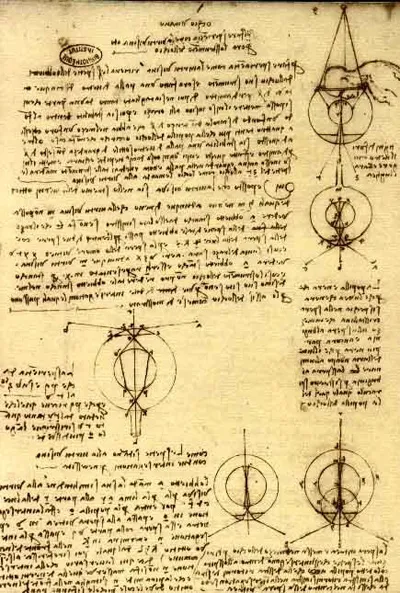
The da Vinci Codex.

Keratoconus described by Nottingham.

Modern view of keratoconus.
In 1801, Thomas Young, an English physician and physicist, having studied medicine in London and Edinburgh and physics at Gottingen, identified the cause of astigmatism and published a three-color theory of perception. While a fellow at Cambridge University, he used a microscope lens with wax around the rim that was filled with water and held against the eye in an experiment on accommodation to neutralize the refractive effect of the cornea [8]. Inspired by Thomas Young, George Biddle Airy, British mathematician and astronomer, colluding and co-operating with John Herschel at Cambridge University, experimented with his own astigmatism. He described not only the optical theory of astigmatism, but also its correction with a theoretical back surface toric lens. He used the first few days of the moon’s crescent (similar to a stenopaic slit) to distinguish the astigmatic axis of hi...
Table of contents
- Welcome
- Table of Contents
- Title Page
- BENTHAM SCIENCE PUBLISHERS LTD.
- FOREWORD
- PREFACE
- List of Contributors
- PROLOGUE
- History of Scleral Lenses
- Scleral Lens Anatomy
- Understanding Anterior Ocular Surface Shape
- Scleral Lens Optics
- Medical Indications for Scleral Lens Use
- Scleral Lenses for the Regular / Normal / Non-Diseased Cornea
- Instrumentation
- Initial Lens Selection
- Scleral Lens Evaluation
- Documentation
- Examination Flow for Scleral Lens Fitting
- Scleral Lens Complications and Problem Solving
- Scleral Lens Handling
- Scleral Lens Challenges
- Scleral Lens Patient Recruitment
- Collaborative Care of the Scleral Lens Patient: Working with Referring Doctors
Frequently asked questions
Yes, you can cancel anytime from the Subscription tab in your account settings on the Perlego website. Your subscription will stay active until the end of your current billing period. Learn how to cancel your subscription
No, books cannot be downloaded as external files, such as PDFs, for use outside of Perlego. However, you can download books within the Perlego app for offline reading on mobile or tablet. Learn how to download books offline
Perlego offers two plans: Essential and Complete
- Essential is ideal for learners and professionals who enjoy exploring a wide range of subjects. Access the Essential Library with 800,000+ trusted titles and best-sellers across business, personal growth, and the humanities. Includes unlimited reading time and Standard Read Aloud voice.
- Complete: Perfect for advanced learners and researchers needing full, unrestricted access. Unlock 1.5M+ books across hundreds of subjects, including academic and specialized titles. The Complete Plan also includes advanced features like Premium Read Aloud and Research Assistant.
We are an online textbook subscription service, where you can get access to an entire online library for less than the price of a single book per month. With over 1.5 million books across 990+ topics, we’ve got you covered! Learn about our mission
Look out for the read-aloud symbol on your next book to see if you can listen to it. The read-aloud tool reads text aloud for you, highlighting the text as it is being read. You can pause it, speed it up and slow it down. Learn more about Read Aloud
Yes! You can use the Perlego app on both iOS and Android devices to read anytime, anywhere — even offline. Perfect for commutes or when you’re on the go.
Please note we cannot support devices running on iOS 13 and Android 7 or earlier. Learn more about using the app
Please note we cannot support devices running on iOS 13 and Android 7 or earlier. Learn more about using the app
Yes, you can access Contemporary Scleral Lenses: Theory and Application by Melissa Barnett,Lynette K. Johns, Melissa Barnett, Lynette K. Johns in PDF and/or ePUB format, as well as other popular books in Medicine & Opthalmology & Optometry. We have over 1.5 million books available in our catalogue for you to explore.