- English
- ePUB (mobile friendly)
- Available on iOS & Android
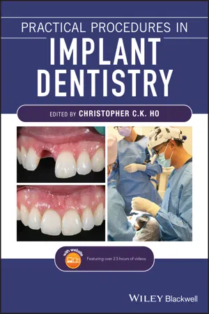
Practical Procedures in Implant Dentistry
About this book
Practical Procedures in IMPLANT DENTISTRY
Master the fundamentals and intricacies of implant dentistry with this comprehensive and practical new resource
Practical Procedures in Implant Dentistry delivers a comprehensive collection of information demonstrating the science and clinical techniques in implant dentistry. Written in a practical and accessible style that outlines the principles and procedures of each technique, the book offers clinical tips and references to build a comprehensive foundation of knowledge in implantology.
Written by an international team of contributors with extensive clinical and academic expertise, Practical Procedures in Implant Dentistry covers core topics such as:
- Rationale and assessment for implant placement and restoration, including the diagnostic records and surgical considerations required for optimal planning and risk management
- Incision design considerations and flap management, with an essential knowledge of regional neuro-vascular structures
- Implant placement, encompassing the timing of the placement, bone requirements and understanding the importance of the peri-implant interface for soft tissue stability
- Impression techniques, loading protocols, digital workflows and the aesthetic considerations of implants
- Prosthetic rehabilitation of single tooth implants to fully edentulous workflows, including discussions of soft tissue support, biomechanics and occlusal verification
Perfect for both general dental practitioners and specialists in implant dentistry, Practical Procedures in Implant Dentistry is also a valuable reference to senior undergraduate and postgraduate dental students.
Tools to learn more effectively

Saving Books

Keyword Search

Annotating Text

Listen to it instead
Information
1
Introduction
- A prosthetically driven approach: Historically, a surgically driven approach was used in which implants were placed in the bony anatomy available. However, in cases of deficiency this resulted in final restorations that were compromised. A prosthetically driven approach is referred to as ‘backwards planning’; the final ideal tooth position is planned, and augmentation may need to be performed to allow the final implant to be in the optimal position.
- Radiographic imaging: Cross‐sectional imaging with cone beam computed tomography (CBCT) scans in combination with the use of planning software allows 3D positioning for the prosthetically planned approach. Improved safety and predictability in implant insertion has resulted. The use of surgically guided templates to provide precise implant placement with alignment in the correct axis enhances predictability and reliability in placing implants that are bodily within bone, and with access alignment that may allow screw retention. It also allows the clinician to diagnose whether augmentation may need to be undertaken in either a simultaneous or staged approach with implant placement.
- Importance of the soft tissue interface: It is now understood that the peri‐implant soft tissues are paramount for long‐term stability and predictability. The soft tissue interface is similar to that of natural teeth and a barrier to microbial invasion. Histologically, peri‐implant tissues possess a junctional epithelium and supracrestal zone of connective tissue. This connective tissue helps seal off the oral environment, with the fibres arranged parallel to the implant surface in a cuff‐like circular orientation. This arrangement may impact how the tissue responds to bacterial insult or cement extrusion into the sulcus. Natural teeth have gingival fibres inserting into cementum tissues, but because of the parallel arrangement of fibres around implants the tissues are more easily detached from the implant surface. This may lead to breakdown such as that seen in peri‐implantitis or cement extrusion. This inflammatory breakdown is often seen at an accelerated rate compared to that of periodontitis. Literature has also demonstrated the presence of a ‘biologic width’ around dental implants, and understanding the influence of thick tissue will help prevent bone loss and provide improved stability [2, 3].
- Implant design: Both macrostructure and microstructure of implants have undergone continuous development to attain better primary stability, quicker osseointegration, and increased bone–implant contact. Micromotion may disturb tissue healing and vasculature, with micromotion greater than 100–150 μm detaching the fibrin clot from the implant surface. Modern implant designs have focused on achieving enhanced primary stability, with manufacturers developing a tapered implant that allows for the widest part of the implant to engage the cortical bone at the crest, while the apical portion is tapered to allow the trabecular bone to be compressed. The original implant connections were an external hex, however modern implant designs have focused on platform‐switched internal connections. These are often conical connections, with several manufacturers’ designs approaching a Morse taper. This creates significant friction through the high degree of parallelism between the two structures within the connection. It has been shown to reduce the microgap size and distribute stress more evenly; there is also increasing evidence that it helps to preserve peri‐implant bone and stabilise soft tissues. Extensive research into implant microstructure has established the optimal environment for bone–implant contact, with both additive and subtractive techniques used to develop moderately rough surfaces (Sa 1–2 μm). Most implant manufacturers produce this surface by using acid etching, grit blasting, or anodic oxidation. This roughness improves the osteoconductivity of the surface.
- Digital implant dentistry – computer‐aided design ( CAD ), computer‐aided manufacturing ( CAM ), chairside intra‐oral scanning, and 3D printing: This area has undergone significant technological improvements in recent years, with implant planning software allowing accurate planning of dental implants using CBCT. The ability to print surgical guides through 3D printing is now commonplace, with many dental practices able to access this technology due to the reduced cost of printing. CAD/CAM fabrication of prosthetic abutments and implant bars allows customised designs that are passively fitting, economic, and homogeneous, with no distortion compared to that of cast metal frameworks. The many different materials dental clinicians have available to mill nowadays, including zirconia, ceramics, hybrid ceramics, cobalt‐chrome, and titanium, allow the modern clinician to select appropriate materials for both aesthetics and strength when required.
- Loading protocols: The original protocols demanded an unloaded period of healing after implant surgery that ranged from three to six months. With the improved designs possessing better primary stability and roughened surface implants, these delayed loading protocols have been challenged, with immediate loading of implants providing immediate function in the first 48 hours. This has led to better acceptance of treatment, with reduced numbers of appointments and intervention. Survival rates are high for immediate loaded and conventional loaded implants, however immediate loading may pose a greater risk for implant failure if there is the possibility of micromotion.
- Complications and long‐term maintenance: Because the original implant patients were treated over 50 years ago now, many patients have had implants for multiple decades. Complications are known. These can be mechanical in nature, such as screw loosening/fracture, veneering material fractures and wear, or biological complications with peri‐implantitis and inflammation. Proper planning minimises such failure and complications. Patients should still understand that regular continuing care is required and that their implant treatment may require servicing and may even need replacement in the future.
References
- 1 Brånemark, P.‐I., Hansson, B.O., Adell, R. et al. (1977). Osseointegrated Implants in the Treatment of the Edentulous Jaw, 132. Stockholm: Almqvist and Wiksell.
- 2 Linkevicius, T., Apse, P., Grybauskas, S., and Puisys, A. (2009). The influence of soft tissue thic...
Table of contents
- Cover
- Table of Contents
- Title Page
- Copyright Page
- Foreword
- List of Contributors
- About the Companion Website
- 1 Introduction
- 2 Patient Assessment and History Taking
- 3 Diagnostic Records
- 4 Medico‐Legal Considerations and Risk Management
- 5 Considerations for Implant Placement
- 6 Anatomic and Biological Principles for Implant Placement
- 7 Maxillary Anatomical Structures
- 8 Mandibular Anatomical Structures
- 9 Extraction Ridge Management
- 10 Implant Materials, Designs, and Surfaces
- 11 Timing of Implant Placement
- 12 Implant Site Preparation
- 13 Loading Protocols in Implantology
- 14 Surgical Instrumentation
- 15 Flap Design and Management for Implant Placement
- 16 Suturing Techniques
- 17 Pre‐surgical Tissue Evaluation and Considerations in Aesthetic Implant Dentistry
- 18 Surgical Protocols for Implant Placement
- 19 Optimising the Peri‐implant Emergence Profile
- 20 Soft Tissue Augmentation
- 21 Bone Augmentation Procedures
- 22 Impression Taking in Implant Dentistry
- 23 Implant Treatment in the Aesthetic Zone
- 24 The Use of Provisionalisation in Implantology
- 25 Abutment Selection
- 26 Screw versus Cemented Implant‐Supported Restorations
- 27 A Laboratory Perspective on Implant Dentistry
- 28 Implant Biomechanics
- 29 Delivering the Definitive Prosthesis
- 30 Occlusion and Implants
- 31 Dental Implant Screw Mechanics
- 32 Prosthodontic Rehabilitation for the Fully Edentulous Patient
- 33 Implant Maintenance
- 34 The Digital Workflow in Implant Dentistry
- 35 Biological Complications
- 36 Implant Prosthetic Complications
- Index
- End User License Agreement
Frequently asked questions
- Essential is ideal for learners and professionals who enjoy exploring a wide range of subjects. Access the Essential Library with 800,000+ trusted titles and best-sellers across business, personal growth, and the humanities. Includes unlimited reading time and Standard Read Aloud voice.
- Complete: Perfect for advanced learners and researchers needing full, unrestricted access. Unlock 1.4M+ books across hundreds of subjects, including academic and specialized titles. The Complete Plan also includes advanced features like Premium Read Aloud and Research Assistant.
Please note we cannot support devices running on iOS 13 and Android 7 or earlier. Learn more about using the app