- English
- ePUB (mobile friendly)
- Available on iOS & Android
eBook - ePub
Brain Disorders Sourcebook, 6th Ed.
About this book
Provides basic consumer health information about the diagnosis and treatment of acquired and traumatic brain injuries, brain disorders, and degenerative diseases and conditions. Includes index, glossary of related terms, directory of resources.
Tools to learn more effectively

Saving Books

Keyword Search

Annotating Text

Listen to it instead
Information
Topic
MédecineSubtopic
Santé généralePart 1 | Brain Basics
Chapter 1 | Know Your Brain
The brain is the most complex part of the human body. This three-pound organ is the seat of intelligence, interpreter of the senses, initiator of body movement, and controller of behavior. Lying in its bony shell and washed by protective fluid, the brain is the source of all the qualities that define our humanity. The brain is the crown jewel of the human body.
For centuries, scientists and philosophers have been fascinated by the brain, but until recently they viewed the brain as nearly incomprehensible. Now, however, the brain is beginning to relinquish its secrets. Scientists have learned more about the brain in the last 10 years than in all previous centuries because of the accelerating pace of research in neurological and behavioral science and the development of new research techniques. As a result, Congress named the 1990s the Decade of the Brain.
This chapter is a basic introduction to the human brain. It may help you understand how the healthy brain works, how to keep it healthy, and what happens when the brain is diseased or dysfunctional.
The Architecture of the Brain
The brain is like a committee of experts. All the parts of the brain work together, but each part has its own special properties. The brain can be divided into three basic units: the forebrain, the midbrain, and the hindbrain.

Figure 1.1. Know Your Brain
(Source: “Brain,” Surveillance, Epidemiology, and End Results (SEER)
Program, National Cancer Institute (NCI).)
(Source: “Brain,” Surveillance, Epidemiology, and End Results (SEER)
Program, National Cancer Institute (NCI).)
The hindbrain includes the upper part of the spinal cord, the brain stem, and a wrinkled ball of tissue called the “cerebellum.” The hindbrain controls the body’s vital functions such as respiration and heart rate. The cerebellum coordinates movement and is involved in learned rote movements. When you play the piano or hit a tennis ball you are activating the cerebellum. The uppermost part of the brainstem is the midbrain, which controls some reflex actions and is part of the circuit involved in the control of eye movements and other voluntary movements. The forebrain is the largest and most highly developed part of the human brain: it consists primarily of the cerebrum and the structures hidden beneath it.
When people see pictures of the brain it is usually the cerebrum that they notice. The cerebrum sits at the topmost part of the brain and is the source of intellectual activities. It holds your memories, allows you to plan, enables you to imagine and think. It allows you to recognize friends, read books, and play games.
The cerebrum is split into two halves (hemispheres) by a deep fissure. Despite the split, the two cerebral hemispheres communicate with each other through a thick tract of nerve fibers that lies at the base of this fissure. Although the two hemispheres seem to be mirror images of each other, they are different. For instance, the ability to form words seems to lie primarily in the left hemisphere, while the right hemisphere seems to control many abstract reasoning skills.
For some as-yet-unknown reason, nearly all of the signals from the brain to the body and vice-versa cross over on their way to and from the brain. This means that the right cerebral hemisphere primarily controls the left side of the body and the left hemisphere primarily controls the right side. When one side of the brain is damaged, the opposite side of the body is affected. For example, a stroke in the right hemisphere of the brain can leave the left arm and leg paralyzed.
The Geography of Thought
Each cerebral hemisphere can be divided into sections, or lobes, each of which specializes in different functions. To understand each lobe and its specialty we will take a tour of the cerebral hemispheres, starting with the two frontal lobes, which lie directly behind the forehead. When you plan a schedule, imagine the future, or use reasoned arguments, these two lobes do much of the work. One of the ways the frontal lobes seem to do these things is by acting as short-term storage sites, allowing one idea to be kept in mind while other ideas are considered. In the rearmost portion of each frontal lobe is a motor area, which helps control voluntary movement. A nearby place on the left frontal lobe called “Broca’s area” allows thoughts to be transformed into words.
When you enjoy a good meal – the taste, aroma, and texture of the food – two sections behind the frontal lobes called the “parietal lobes” are at work. The forward parts of these lobes, just behind the motor areas, are the primary sensory areas. These areas receive information about temperature, taste, touch, and movement from the rest of the body. Reading and arithmetic are also functions in the repertoire of each parietal lobe.
As you look at the words and pictures on this page, two areas at the back of the brain are at work. These lobes, called the “occipital lobes,” process images from the eyes and link that information

Figure 1.2. The Forebrain
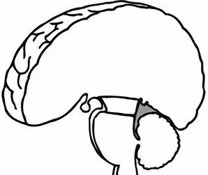
Figure 1.3. The Midbrain

Figure 1.4. The Hindbrain
with images stored in memory. Damage to the occipital lobes can cause blindness.
The last lobes on our tour of the cerebral hemispheres are the temporal lobes, which lie in front of the visual areas and nest under the parietal and frontal lobes. Whether you appreciate symphonies or rock music, your brain responds through the activity of these lobes. At the top of each temporal lobe is an area responsible for receiving information from the ears. The underside of each temporal lobe plays a crucial role in forming and retrieving memories, including those associated with music. Other parts of this lobe seem to integrate memories and sensations of taste, sound, sight, and touch.
The Cerebral Cortex
Coating the surface of the cerebrum and the cerebellum is a vital layer of tissue the thickness of a stack of two or three dimes. It is called the “cortex,” from the Latin word for bark. Most of the actual information processing in the brain takes place in the cerebral cortex. When people talk about “gray matter” in the brain they are talking about this thin rind. The cortex is gray because nerves in this area lack the insulation that makes most other parts of the brain appear to be white. The folds in the brain add to its surface area and therefore increase the amount of gray matter and the quantity of information that can be processed.
The Inner Brain
Deep within the brain, hidden from view, lie structures that are the gatekeepers between the spinal cord and the cerebral hemispheres. These structures not only determine our emotional state, they also modify our perceptions and responses depending on that state, and allow us to initiate movements that you make without thinking about them. Like the lobes in the cerebral hemispheres, the structures described below come in pairs: each is duplicated in the opposite half of the brain.
The hypothalamus, about the size of a pearl, directs a multitude of important functions. It wakes you up in the morning, and gets

Figure 1.5. Inner Brain
the adrenaline flowing during a test or job interview. The hypothalamus is also an important emotional center, controlling the molecules that make you feel exhilarated, angry, or unhappy. Near the hypothalamus lies the thalamus, a major clearinghouse for information going to and from the spinal cord and the cerebrum.
An arching tract of nerve cells leads from the hypothalamus and the thalamus to the hippocampus. This tiny nub acts as a memory indexer – sending memories out to the appropriate part of the cerebral hemisphere for long-term storage and retrieving them when necessary. The basal ganglia (not shown) are clusters of nerve cells surrounding the thalamus. They are responsible for initiating and integrating movements. Parkinson disease, which results in tremors, rigidity, and a stiff, shuffling walk, is a disease of nerve cells that lead into the basal ganglia.
Making Connections
The brain and the rest of the nervous system are composed of many different types of cells, but the primary functional unit is a cell called “the neuron.” All sensations, movements, thoughts, memories, and feelings are the result of signals that pass through neurons. Neurons consist of three parts. The cell body contains

Figure 1.6. A Typical Neuron
the nucleus, where most of the molecules that the neuron needs to survive and function are manufactured. Dendrites extend out from the cell body like the branches of a tree and receive messages from other nerve cells. Signals then pass from the dendrites through the cell body and may travel away from the cell body down an axon to another neuron, a muscle cell, or cells in some other organ. The neuron is usually surrounded by many support cells. Some types of cells wrap around the axon to form an insulating sheath. This sheath can include a fatty molecule called “myelin,” which provides insulation for the axon and helps nerve signals travel faster and farther. Axons may be very short, such as those that carry signals from one cell in the cortex to another cell less than a hair’s width away. Or axons may be very long, such as those that carry messages from the brain all the way down the spinal cord.
Scientists have learned a great deal about neurons by studying the synapse – the place where a signal passes from the neuron to another cell. When the signal reaches the end of the axon it stimulates the release of tiny sacs. These sacs release chemicals known

Figure 1.7. Synapse
as...
Table of contents
- Cover
- Halftitle
- Title
- Copyright
- Table of Contents
- Preface
- Part 1. Brain Basics
- Part 2. Diagnosing and Treating Brain Disorders
- Part 3. Genetic and Congenital Brain Disorders
- Part 4. Brain Infections
- Part 5. Acquired and Traumatic Brain Injuries
- Part 6. Brain Tumors
- Part 7. Degenerative Brain Disorders
- Part 8. Seizures and Neurological Disorders of Sleep
- Part 9. Additional Help and Information
- Index
Frequently asked questions
Yes, you can cancel anytime from the Subscription tab in your account settings on the Perlego website. Your subscription will stay active until the end of your current billing period. Learn how to cancel your subscription
No, books cannot be downloaded as external files, such as PDFs, for use outside of Perlego. However, you can download books within the Perlego app for offline reading on mobile or tablet. Learn how to download books offline
Perlego offers two plans: Essential and Complete
- Essential is ideal for learners and professionals who enjoy exploring a wide range of subjects. Access the Essential Library with 800,000+ trusted titles and best-sellers across business, personal growth, and the humanities. Includes unlimited reading time and Standard Read Aloud voice.
- Complete: Perfect for advanced learners and researchers needing full, unrestricted access. Unlock 1.4M+ books across hundreds of subjects, including academic and specialized titles. The Complete Plan also includes advanced features like Premium Read Aloud and Research Assistant.
We are an online textbook subscription service, where you can get access to an entire online library for less than the price of a single book per month. With over 1 million books across 990+ topics, we’ve got you covered! Learn about our mission
Look out for the read-aloud symbol on your next book to see if you can listen to it. The read-aloud tool reads text aloud for you, highlighting the text as it is being read. You can pause it, speed it up and slow it down. Learn more about Read Aloud
Yes! You can use the Perlego app on both iOS and Android devices to read anytime, anywhere — even offline. Perfect for commutes or when you’re on the go.
Please note we cannot support devices running on iOS 13 and Android 7 or earlier. Learn more about using the app
Please note we cannot support devices running on iOS 13 and Android 7 or earlier. Learn more about using the app
Yes, you can access Brain Disorders Sourcebook, 6th Ed. by James Chambers in PDF and/or ePUB format, as well as other popular books in Médecine & Santé générale. We have over one million books available in our catalogue for you to explore.